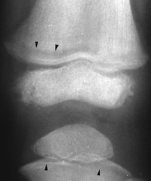
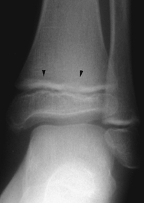
CASE 98 A lethargic 7-year-old boy presents with painful knees/shins. Figure 98A Figure 98B Coned-down frontal views centered on the knee and ankle demonstrates subphyseal metaphyseal lucent bands involving distal femur as well as proximal and distal tibia (Figs. 98A and 98B). The bone configuration/modeling appears normal. Acute lymphoblastic leukemia (ALL) The differential diagnosis of lucent metaphyseal bands are: Of all pediatric malignancies, leukemia is the most common, accounting for 30 to 40% of all childhood cancers. ALL is the most common form, comprising up to 85% of patients. The disease is most frequent in children 2 to 5 years of age. Skeletal manifestations may be the initial presentation in up to 20% of cases. Between 70 and 90% of children with leukemia will manifest skeletal radiographic changes during their course of illness and treatment. Long bones (usually lower limbs) are more often affected than axial bones. Radiation exposure has been linked with the development of ALL; patients with ataxia telangiectasia, Down syndrome, and Fanconi’s anemia are also predisposed. Oncogenic mechanisms include enhanced expression of protooncogenes (MYC, TAL-1, LYL-1, HOX-11), the expression of translocation-generated fusion oncogenes (BCR-ABL, TEL-AML-1, E2A-PBX-1, and MLL fusions), and alterations in chromosomal number (decrease or increase). The majority of these mechanisms are insufficient on their own to generate a full leukemic phenotype. The nature and number of mutations needed to induce leukemia appear to vary, depending on unknown initiation lesions, some of which may be acquired in utero. Figure 98C Frontal knee radiograph depicts subphyseal distal femoral metaphyseal lucent band paralleling growth plate. Similar, less prominent lucency spans subphyseal metaphysis of proximal tibia (arrowheads). Figure 98D
Clinical Presentation
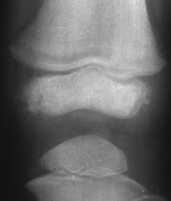
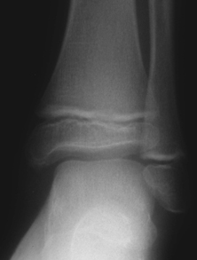
Radiologic Findings
Diagnosis
Differential Diagnosis
Discussion
Background
Etiology
Clinical Findings
Complications
![]()
Stay updated, free articles. Join our Telegram channel


Full access? Get Clinical Tree


