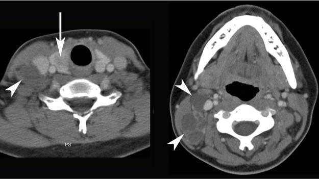
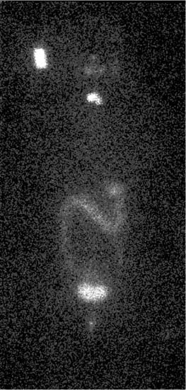
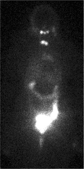
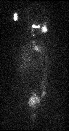
CASE 99 A 24-year-old woman presents with palpable right-sided neck nodes. A CT scan is requested for further assessment. Fig. 99.1 Fig. 99.2 Initial whole body scan. Fig. 99.3 Post-therapy scan. Fig. 99.4 Three years later: whole body scan. • 131I, 2 mCi (74 MBq) orally. Planar imaging with a high-energy collimator 2 days later (Figs. 99.2 and 99.4). • 131I, 150 mCi (5550 MBq) orally (therapy dose). Planar imaging with a high-energy collimator 7 days later (Fig. 99.3). CT images at two levels through the neck (Fig. 99.1) demonstrate multiple rim-enhancing enlarged lymph nodes (arrowheads), many with cystic centers. A low-density lesion is also seen in the right lobe of the thyroid (arrow). This is biopsied and shown to be papillary thyroid cancer. Following thyroidectomy, a 131I scan is performed (Fig. 99.2) in preparation for radioiodine therapy. This demonstrates uptake in the thyroid remnants, with no metastases identified. A rectangular area of activity to the right of the head (Figs. 99.2 and 99.4) is within a standard of known activity placed next to the patient for uptake calcification purposes. The patient was administered an oral dose of 150 mCi of 131I for thyroid ablation and adjuvant therapy. A post-therapy scan 1 week later (Fig. 99.3
Clinical Presentation
Technique
Image Interpretation
Therapy
![]()
Stay updated, free articles. Join our Telegram channel


Full access? Get Clinical Tree


