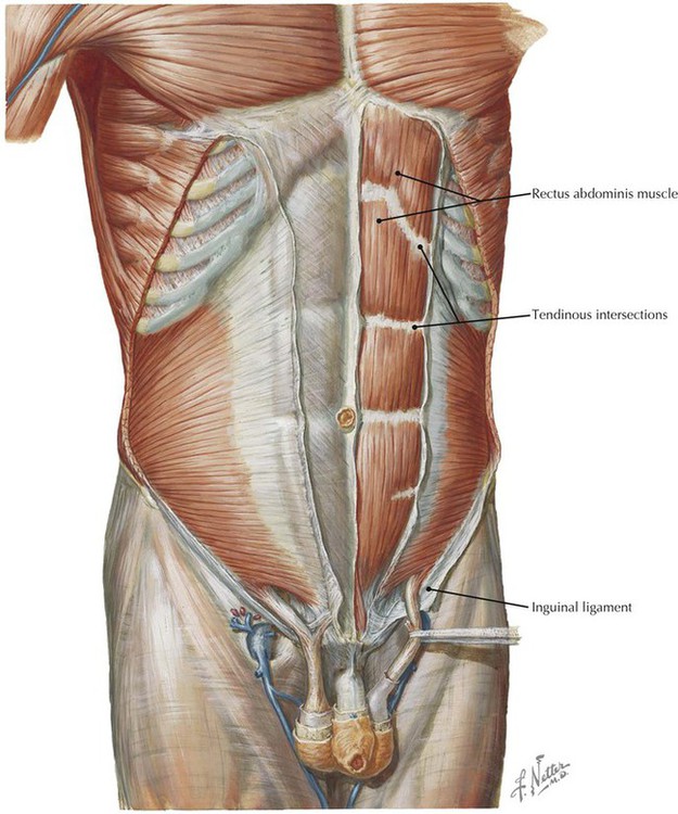
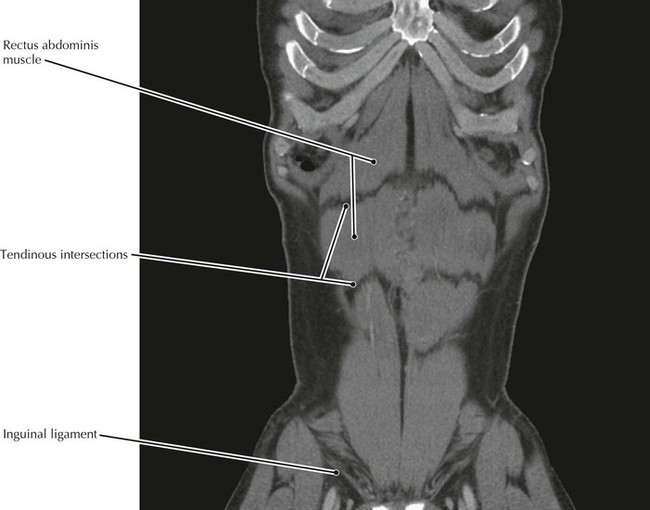
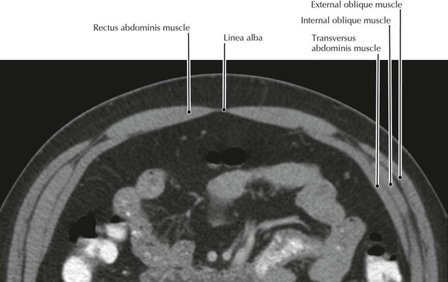
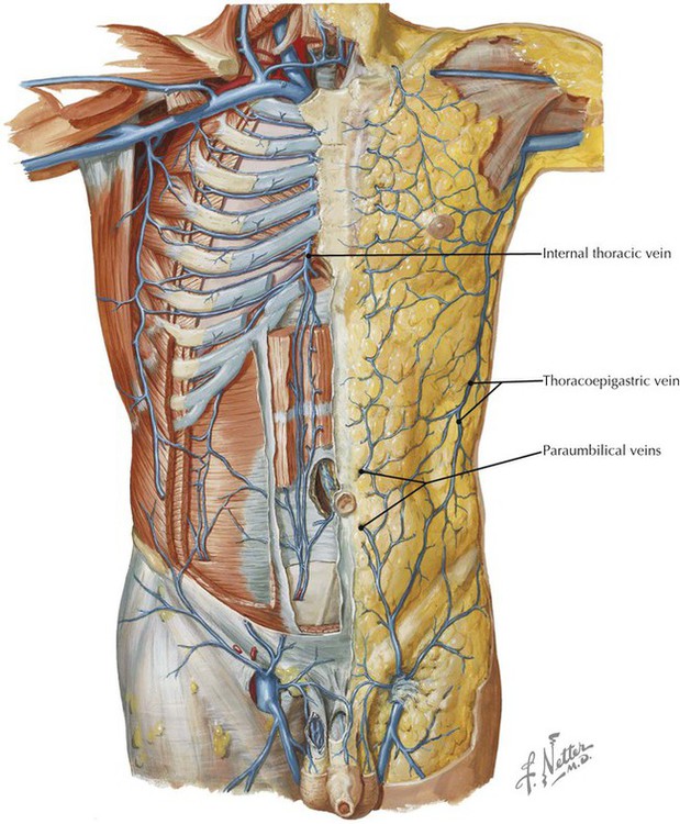
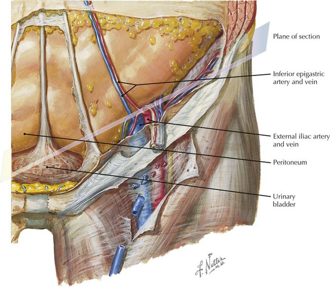
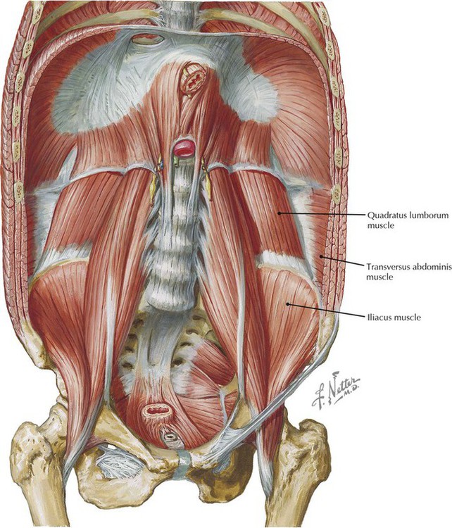
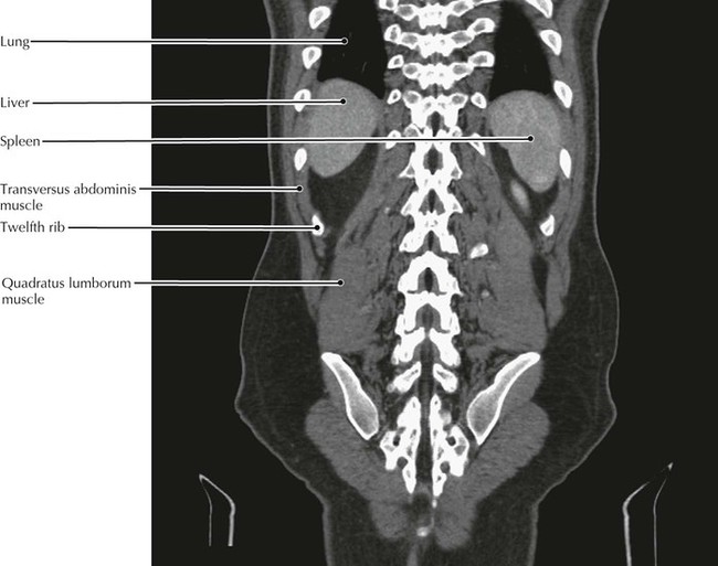
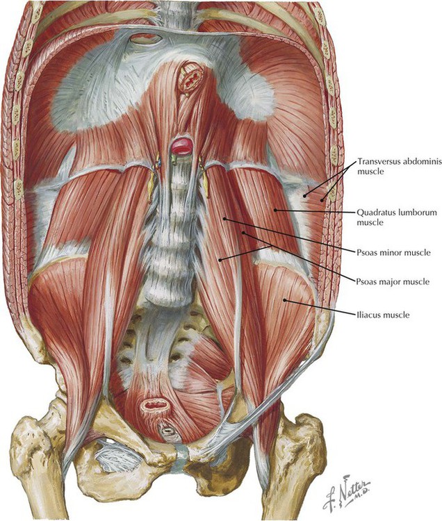
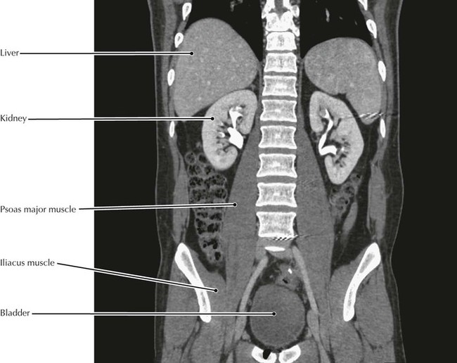
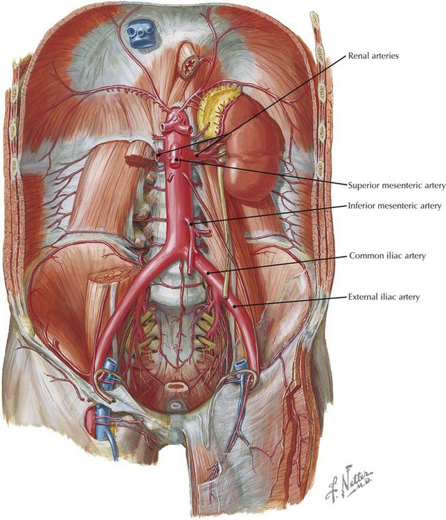
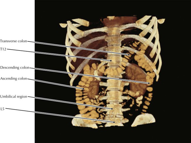
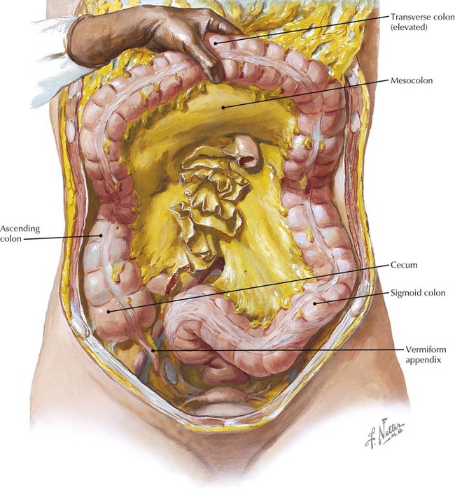
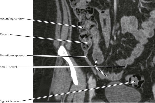
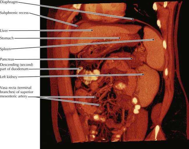
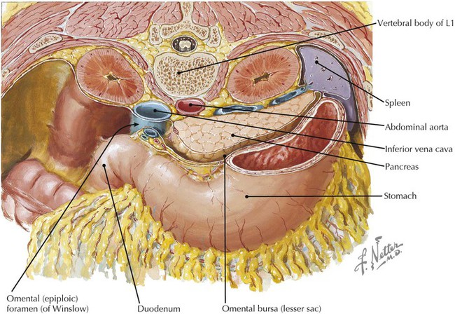
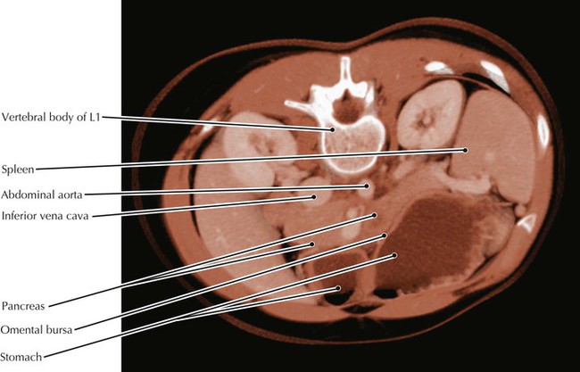
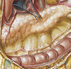
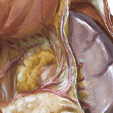
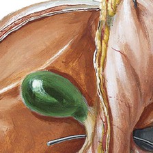
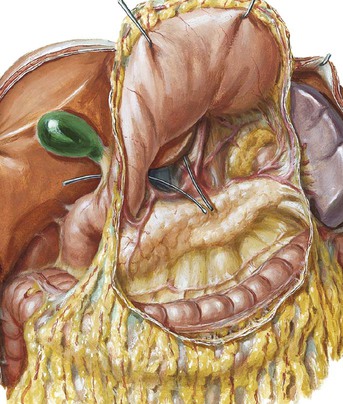
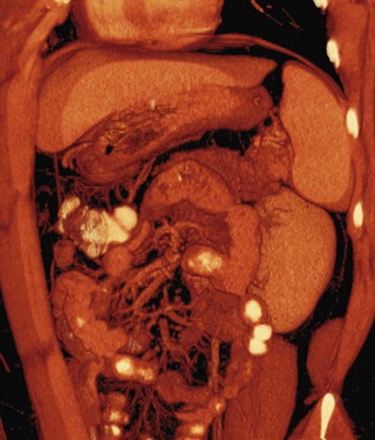
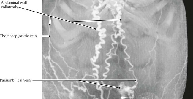
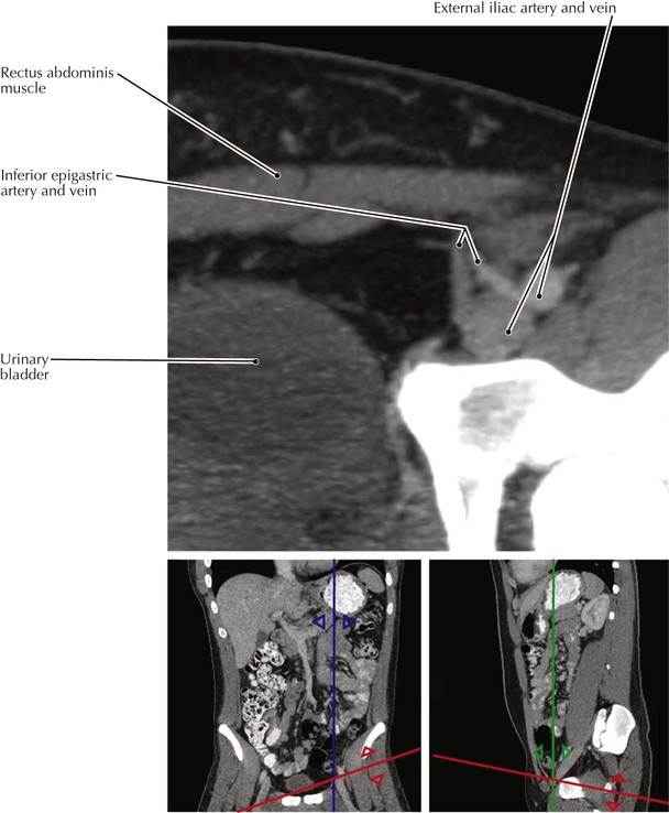
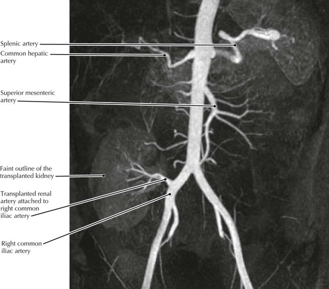
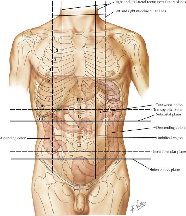
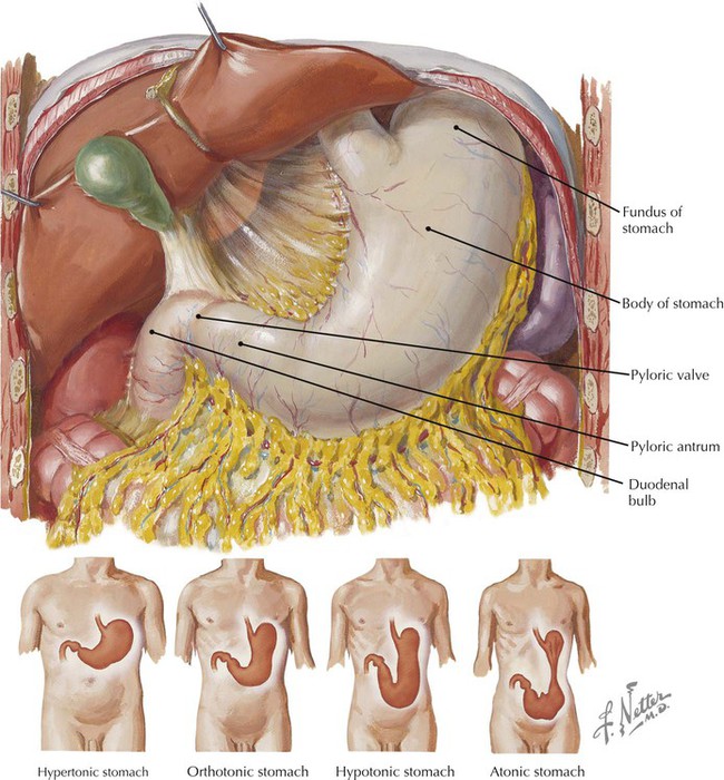
• The linea alba is composed of the interweaving fibers of the aponeuroses of the abdominal muscles and is important surgically because longitudinal incisions in it are relatively bloodless. • The composition of the anterior and posterior layers of the rectus sheath changes superior and inferior to the arcuate line (of Douglas), which is where the inferior epigastric artery enters the sheath. • Abdominal wall collaterals join the internal thoracic (mammary) and lateral thoracic veins to return venous blood to the vena cava. • The paraumbilical veins communicate with the portal vein via the vein in the ligamentum teres hepatis (round ligament of the liver). • When pathology obstructs normal flow, collateral vessels may dilate and become tortuous as shown in this CT. • The inferior epigastric vessels are an important landmark for differentiating between indirect and direct inguinal hernias. Pulsations from the artery can be felt medial to the neck of an indirect hernia and lateral to the neck of a direct hernia. • The inferior epigastric vessels enter the rectus sheath approximately at the arcuate line, which is where the formation of the sheath changes. Inferior to the line the aponeuroses of all of the abdominal muscles pass anterior to the rectus abdominis muscle whereas superior to the line, half of the aponeurosis of the internal oblique muscle and all of the aponeurosis of the transversus abdominis pass posterior to the rectus muscle. • The quadratus lumborum muscle primarily laterally flexes the trunk when acting unilaterally. • The quadratus lumborum muscle attaches to the 12th rib and thereby can act as an accessory respiratory muscle by allowing the diaphragm to exert greater downward force by preventing upward movement of the 12th rib. • Patency of the anastomosis (connection) of the iliac artery to the transplanted renal artery is demonstrated. • The indication for kidney transplantation is end-stage renal disease (ESRD). Diabetes is the most common cause of ESRD, followed by glomerulonephritis. • Potential recipients of kidney transplants undergo an extensive immunologic evaluation to minimize transplants that are at risk for antibody-mediated hyperacute rejection. • The left kidney is the one preferred for transplant because of its longer vein compared to the right. • Classically, the abdomen is divided into four quadrants defined by vertical and horizontal planes through the umbilicus. More recently, it has been divided into nine regions based on subcostal, transtubercular, and right and left lateral rectus (semilunar) planes. • Note the greater height of the left colic (splenic) flexure compared to the hepatic flexure on the right. • Inspissated bowel contents may lead to development of an appendolith, which is a calcified concretion that may obstruct the proximal lumen of the appendix; stasis, bacterial overgrowth, infection, and swelling (i.e., appendicitis) may follow, as can eventual rupture. • The appendix is highly variable in its location, including occasionally being posterior to the cecum (retrocecal). • The right kidney is not apparent in this image because of the obliquity of the image (the plane of the “coronal” image is angled so that it passes anterior to the right kidney but through the left kidney). • The vasa recta (terminal branches) of the superior mesenteric artery (SMA) supply loops of small bowel. • The terminal or fourth segment of the duodenum is attached to the diaphragm by a variable band of smooth muscle known as the suspensory ligament of the duodenum (ligament of Treitz). It is not recognizable on CT images. • The stomach is filled with whole milk in this patient, the fat content of which decreases the CT density of the stomach fluid in order to enhance contrast differences with other tissues, such as the stomach wall. Note that the pyloric valve is closed, as it is most of the time. • The position of the stomach is variable in relation to the body habitus. This patient has an “orthotonic” stomach.
Abdomen
Anterior Abdominal Wall Muscles
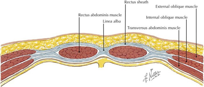
Abdominal Wall, Superficial View
Inguinal Region
Quadratus Lumborum
Kidneys, Normal and Transplanted
Abdominal Regions
Appendix
Abdomen, Upper Viscera
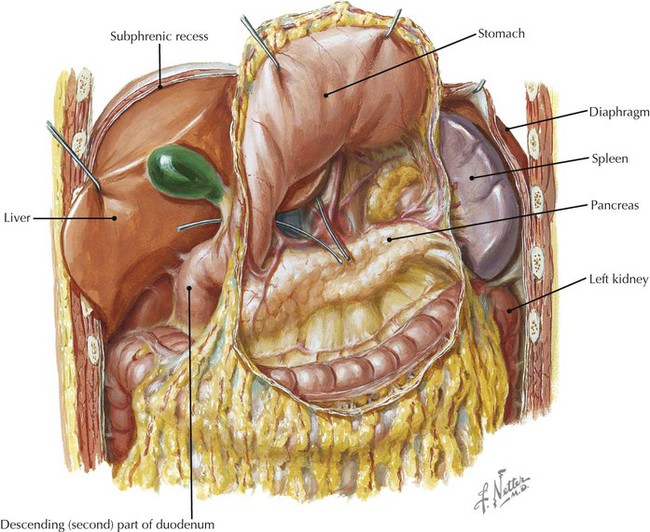
Stomach, In Situ
![]()
Stay updated, free articles. Join our Telegram channel

Full access? Get Clinical Tree


Abdomen

