52 Abdomen: Motion Correction
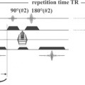
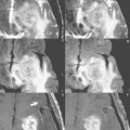
Physiologic motion (e.g., respiration and cardiac pulsation) can lead to artifacts, reducing the diagnostic quality of MR images. Most MR systems offer hardware, software, and sequence-based options designed to minimize or eliminate the effects of physiologic motion during a scan acquisition. Motion artifacts in MR images are caused by translations or shifts in the position of the imaged structure during the acquisition of data. Routine spin echo and gradient echo sequences encode and acquire data to fill k space in a cartesian fashion, with the collection of each line separated by a certain amount of time (TR). In fast spin echo imaging, several, but not all, lines are collected with each TR. Because all the data for an entire slice are not acquired simultaneously, changes in the location of anatomic structures between TR periods can lead to misalignment in spatial encoding. Reconstruction of the data results in blurring or ghosting (see Case 99) in the final image, as illustrated in Fig. 52.1.
The simplest and most effective method for eliminating respiratory motion is to have the patient hold his or her breath. Rapidly acquired T1- and T2-weighted gradient echo sequences are used to collect data during suspended respiration, thereby minimizing motion artifacts. However, this approach requires the scan time to be 25 sec or less for the average patient, leading to limitations in spatial resolution and/or number of slices. The use of this approach is also restricted by the patient’s health and mental status, as well as his or her age. Single-slice techniques such as HASTE (see Case 19), trueFISP (see Case 27
Related posts:

Stay updated, free articles. Join our Telegram channel

Full access? Get Clinical Tree


