Abdominal Wall Hernia
KEY FACTS
Clinical Issues
 Spigelian: Defect in transversus abdominis fascia alongside lateral border of rectus abdominis muscle (linea semilunaris) and lateral border of rectus sheath
 Lumbar: Occur in 2 areas of potential weakness in flank
 Superior lumbar triangle (Grynfeltt hernia) with erector spinae medially, 12th rib superiorly, and internal oblique muscle laterally
 Inferior lumbar triangle (Petit hernia) with latissimus dorsi muscle medially, iliac crest inferiorly, and external oblique muscle laterally
Diagnostic Checklist
IMAGING
General Features
 Abdominal wall; anatomy
 Anterior wall: Paired rectus muscles enclosed by rectus sheath (aponeurosis of internal oblique, external oblique, and transversus abdominis muscles), which laterally forms linea semilunaris and centrally fuses to form linea alba
 Anterolateral-lateral wall: External oblique, internal oblique, and transversus abdominis
 Posterior wall: Quadratus lumborum and erector spinae muscles
 Epigastric hernia
 Umbilical hernia
 Hypogastric hernia
 Spigelian hernia
 Lumbar hernia
 Incisional hernia
Abdominal Wall Hernia


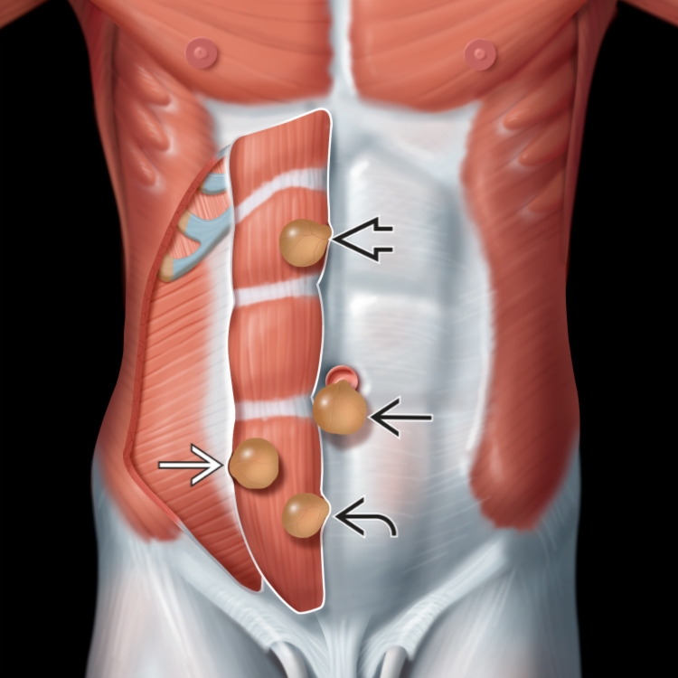
 arising from the umbilicus area. The location of epigastric
  , spigelian
  , and hypogastric
  hernias is also shown for reference.
 at the superior aspect of the umbilicus.
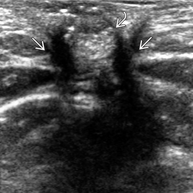
 and no bowel. The margins of the defect
  in the linea alba are clearly seen.
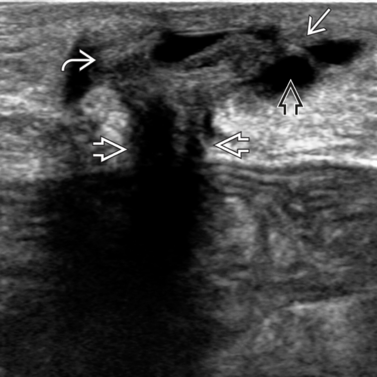
 extending through a defect
  in the linea alba at the umbilicus. The sac contains both echogenic omental fat
  and hypoechoic peritoneal fluid
  . No bowel content is present.