, Marilyn J. Siegel2, Tomasz Miszalski-Jamka3, 4 and Robert Pelberg1
(1)
The Christ Hospital Heart and Vascular Center of Greater Cincinnati, The Lindner Center for Research and Education, Cincinnati, OH, USA
(2)
Mallinckrodt Institute of Radiology, Washington University School of Medicine, St. Louis, Missouri, USA
(3)
Department of Clinical Radiology and Imaging Diagnostics, 4th Military Hospital, Wrocław, Poland
(4)
Center for Diagnosis Prevention and Telemedicine, John Paul II Hospital, Kraków, Poland
Abstract
Univentricular heart, also termed “functionally single ventricle” and “single ventricle,” refers to the congenital heart defects in which the heart functionally has only one pumping chamber. The unifying criteria to identify a univentricular heart is a main (dominant) ventricle with a malformed atrioventricular connection and a rudimentary (incomplete) ventricle, which lacks an inlet component or less commonly an outlet component [1–4]. A true single ventricle is exceedingly rare.
18.1 Univentricular Heart (Double-Inlet Ventricle)
Univentricular heart, also termed “functionally single ventricle” and “single ventricle,” refers to the congenital heart defects in which the heart functionally has only one pumping chamber. The unifying criteria to identify a univentricular heart is a main (dominant) ventricle with a malformed atrioventricular connection and a rudimentary (incomplete) ventricle, which lacks an inlet component or less commonly an outlet component [1–4]. A true single ventricle is exceedingly rare.
The spectrum of univentricular heart anomalies includes absence of one atrioventricular valve (mitral or tricuspid atresia) (see Chap. 12), atresia or severe stenosis of a semilunar valve (aortic atresia, hypoplastic left heart syndrome, and pulmonary atresia with intact ventricular septum) (see Chap. 14), heterotaxy syndrome with only one well-developed ventricle (univentricular heterotaxy syndrome) (see Chap. 20), and double-inlet ventricle (left or right). Double-inlet ventricle is the focus of the discussion in this section.
The well-developed single (dominant) ventricle may demonstrate left, right, or indeterminate morphology. The characteristics of the morphologic left and right ventricles are discussed in Chap. 2. Briefly, the morphologic left ventricle has relatively smooth walls and fine trabeculations and lacks septal chordal attachments to the atrioventricular valve. The morphologic right ventricles is more trabeculated and has septal chordal attachments to its atrioventricular valve [3, 4]. The rare solitary chamber demonstrates coarse apical trabeculation [5]. The atrioventricular connection can be a single (either tricuspid or mitral valve), double, or common inlet (Figs. 18.1 and 18.2).
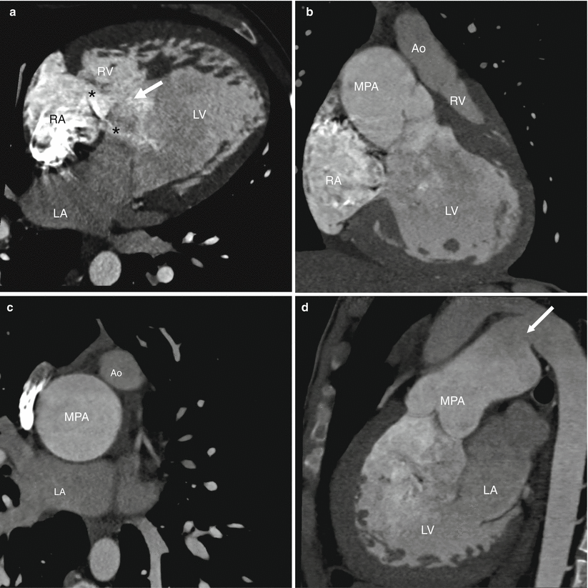
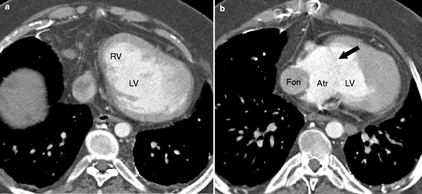
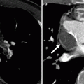
Fig. 18.1
Double-inlet left ventricle with left ventricular morphology and two separate atrioventricular valves and L-transposition of the great arteries, hypoplastic ascending aorta, and persistent ductus arteriosus in a 21-year-old male. Panel (a) is an axial image showing the separate right and left atrioventricular junctions (asterisks) opening into the same dominant chamber with left ventricle (LV) morphology (smooth walls, fine trabeculations). The rudimentary right ventricle (RV) is positioned anteriorly and superiorly. The right atrium (RA) is located anteriorly and to the right, while the left atrium (LA) is positioned posteriorly and to the left, consistent with normal situs. Also note the ventricular septal defect (arrow). Panel (b) is an oblique coronal scan showing discordant ventriculoarterial connections. The dominant LV gives rise to the main pulmonary artery and the rudimentary right ventricle gives rise to a hypoplastic aorta. The aorta lies anteriorly and to the left of the main pulmonary artery (MPA) consistent with L-transposition. Panel (c) is an axial reconstruction again highlighting the hypoplastic aorta (Ao) which is positioned anteriorly and to the left of the pulmonary artery. Panel (d) is a sagittal cut demonstrating the persistent ductus arteriosus (PDA) (arrow) coursing between the main pulmonary artery and the aorta
Fig. 18.2
Double-inlet left ventricle with left ventricular morphology and common atrioventricular valve in an 18-year-old male status post Fontan operation. Panel (a), an axial reconstruction, demonstrates a dominant left ventricle (LV) and a hypoplastic right ventricle (RV) positioned anteriorly. Panel (b), another axial scan reconstructed at a lower plane, shows a common atrium (Atr) with a common atrioventricular valve (arrow) opening into the dominant left ventricular chamber (LV). Fon lateral Fontan tunnel
18.1.1 Double-Inlet Ventricle
This condition is characterized by abnormal atrioventricular connections, with both atria emptying into a single common ventricle (double inlet) that is morphologically predominantly left, predominantly right, or indeterminate (Figs. 18.1, 18.2, and 18.3) [5–9]. When there is a common valve and one well-developed ventricle, the anomaly is also called an unbalanced atrioventricular canal defect (see Chap. 11) [3, 6, 7].
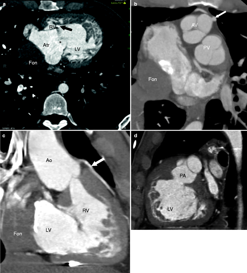
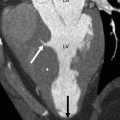
Fig. 18.3
Double-inlet left ventricle with left ventricular morphology, single atrioventricular valve (tricuspid valve atresia), and D-transposition of the great arteries in a 28-year-old male status post Fontan operation. Panel (a) an axial image depicting a rudimentary right ventricle (RV) connected via a small ventricular septal defect (arrow) to the dominant left ventricle (LV). Connection to the common atrium (Atr) is through a single mitral valve (asterisk) since this patient has tricuspid valve atresia. There is no atrioventricular connection to the right ventricular chamber. Note the lateral Fontan circulation (Fon). Panel (b) an axial view showing the aortic valve (AV) with its coronary artery (arrow) anterior and to the right of the pulmonary valve (PV) consistent with D-transposition. The Fontan circulation is also visible. Panel (c) an oblique coronal view illustrating the hypoplastic right ventricle/conus (RV), which gives rise to the aorta (Ao). The Fontan conduit is seen next to the LV. Panel (d) an oblique sagittal image demonstrating the dominant left ventricle (LV) giving rise to the pulmonary artery (PA). White Arrows: coronary artery
Double-inlet ventricle is a rare cardiac malformation accounting for approximately 1.5 % of congenital heart diseases [8].
A comprehensive description of morphology should include (a) ventricle morphology (left, right, indeterminate), (b) atrioventricular valve connections (common, single, or two separate valves), (c) interventricular communication (unrestrictive or restrictive ventricular septal defect), (d) ventriculoarterial connections (concordant, discordant/transposition of great arteries, double-outlet configuration, and common arterial trunk), and (e) atriovisceral situs (solitus, inversus, isomerism) [3–5, 7, 8].
A double-inlet left ventricle has dominant left ventricular morphology and a rudimentary right ventricle located anterosuperiorly (Figs. 18.1, 18.2, and 18.3). A double-inlet right ventricle has dominant right ventricular morphology and a rudimentary left ventricle positioned posteroinferiorly (Fig. 18.4). Double-inlet left ventricle is the more common arrangement [5, 6]. A single ventricular chamber, which is rare, has indeterminate morphology.
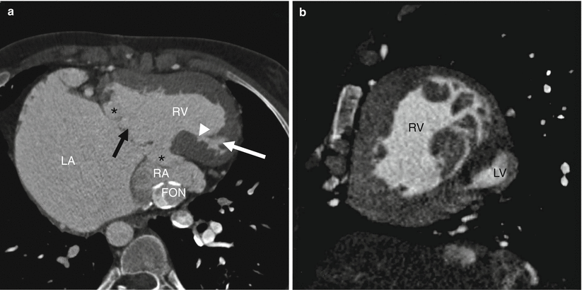
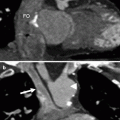
Fig. 18.4
Double-inlet right ventricle with situs inversus in a 21-year-old male after Fontan procedure. Panel (a) an axial image showing that both atrioventricular junctions (asterisks) are supported by the same dominant ventricular chamber of right ventricle (RV) morphology. The rudimentary left ventricle (white arrow) is positioned posteriorly. Note the ventricular septal defect (white arrowhead). The morphological right atrium (RA) is located posteriorly and to the left, while the morphological left atrium (LA) is positioned anteriorly and to the right consistent with situs inversus. Panel (b) a sagittal view showing the dominant ventricle with coarse trabeculations typical of right ventricular morphology (RV) and hypoplastic left ventricle (LV). Black arrow in panel (a): common atrioventricular valve. Fon Fontan circulation
The term double-inlet single ventricle indicates that more than 50 % of the time, both atrioventricular valves or a single common atrioventricular valve opens into one ventricular chamber. There may be a common, single, or double connection. With double-inlet connections, morphological features of the atrioventricular valve may not be distinct enough to distinguish between mitral and tricuspid configurations and the valves are better described as right- or left-sided valves.
The main and rudimentary chambers are separated by a septum, which does not extend to the crux of the heart and is connected to the dominant chamber via a ventricular septal defect (VSD). The VSD is often the muscular-type.
The VSD may be unrestrictive or restrictive initially or become restrictive over time [5]. A restrictive ventricular septal defect leads effectively to subaortic stenosis, which can increase pulmonary blood flow. Ventricular septal defect in a double-inlet ventricle should be differentiated from a true ventricular septal defect. In the latter setting, the septal defect is associated with normally developed right and left ventricular cavities [5].
The great arteries may be normally related (also called “Holmes heart”) or the aorta may be anterior and rightward (D-transposition) or leftward (L-transposition) or “inverted” with a posterior and leftward orientation. In double-inlet left ventricle, the ventriculoarterial connections are generally discordant with the aorta arising from the hypoplastic right ventricle and lying anterior and to the left of the left ventricle (L-transposition). Concordant connections or common arterial trunk configuration is less common [3–5, 10]. In double-inlet right ventricle morphology, both great arteries usually arise from the right ventricle (double-outlet arrangement) or they may have a concordant arrangement. Discordant connections and common arterial trunk are extremely rare in right ventricular morphology [3, 4


Related posts:


Stay updated, free articles. Join our Telegram channel
Full access? Get Clinical Tree
