, Marilyn J. Siegel2, Tomasz Miszalski-Jamka3, 4 and Robert Pelberg1
(1)
The Christ Hospital Heart and Vascular Center of Greater Cincinnati, The Lindner Center for Research and Education, Cincinnati, OH, USA
(2)
Mallinckrodt Institute of Radiology, Washington University School of Medicine, St. Louis, Missouri, USA
(3)
Department of Clinical Radiology and Imaging Diagnostics, 4th Military Hospital, Wrocław, Poland
(4)
Center for Diagnosis Prevention and Telemedicine, John Paul II Hospital, Kraków, Poland
Abstract
In 1969, Rastelli and colleagues introduced a procedure that bypasses left ventricular outflow tract obstructions (LVOT) in complex congenital heart disease patients [1, 2]. In the first step of the Rastelli procedure, the pulmonary artery is separated from the morphologic left ventricle just above the valve, and the cardiac end of the left ventricular outflow tract is closed. Next, an intraventricular tunnel is created using a patch to redirect blood from the left ventricle across the ventricular septal defect (VSD) into the right ventricle and out into the ascending aorta. Then, the right ventricle is anastomosed to the pulmonary artery with an extracardiac conduit, usually using a pulmonary allograft. In the setting of transposition of the great vessels, this procedure corrects the abnormal blood flow pattern at the ventricular level and allows the left ventricle to function as the systemic ventricle [3].
In 1969, Rastelli and colleagues introduced a procedure that bypasses left ventricular outflow tract obstructions (LVOT) in complex congenital heart disease patients [1, 2]. In the first step of the Rastelli procedure, the pulmonary artery is separated from the morphologic left ventricle just above the valve, and the cardiac end of the left ventricular outflow tract is closed. Next, an intraventricular tunnel is created using a patch to redirect blood from the left ventricle across the ventricular septal defect (VSD) into the right ventricle and out into the ascending aorta. Then, the right ventricle is anastomosed to the pulmonary artery with an extracardiac conduit, usually using a pulmonary allograft. In the setting of transposition of the great vessels, this procedure corrects the abnormal blood flow pattern at the ventricular level and allows the left ventricle to function as the systemic ventricle [3].
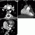
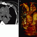
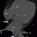
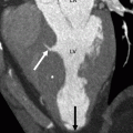
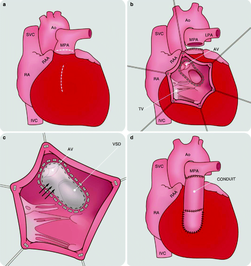
Figures 26.1 and 26.2 illustrate the Rastelli procedure.
Get Clinical Tree app for offline access

Fig. 26.1
An artist’s rendition of the Rastelli procedure. In panel (a) the right ventricle free wall and main pulmonary artery (MPA) are incised (dashed lines). In panel (b) the left ventricle to MPA connection is transected and sutured. Panel (c) shows the creation of an intraventricular tunnel or baffle (arrows) is created to establish a communication between left ventricle and aorta. The VSD and aortic annulus are incorporated within the right ventricle. This step redirects blood from the left ventricle into the right ventricle and out the ascending aorta. Finally, in panel (d), the right ventricle is connected to MPA via a valved conduit. Ao aorta, AV aortic valve, LPA left pulmonary artery, IVC


Stay updated, free articles. Join our Telegram channel

Full access? Get Clinical Tree


