, Marilyn J. Siegel2, Tomasz Miszalski-Jamka3, 4 and Robert Pelberg1
(1)
The Christ Hospital Heart and Vascular Center of Greater Cincinnati, The Lindner Center for Research and Education, Cincinnati, OH, USA
(2)
Mallinckrodt Institute of Radiology, Washington University School of Medicine, St. Louis, Missouri, USA
(3)
Department of Clinical Radiology and Imaging Diagnostics, 4th Military Hospital, Wrocław, Poland
(4)
Center for Diagnosis Prevention and Telemedicine, John Paul II Hospital, Kraków, Poland
Abstract
Arrhythmogenic right ventricular dysplasia (ARVD) is a nonischemic, genetically determined, myocardial disease that involves primarily the right ventricle (RV) and is associated with life-threatening ventricular arrhythmias. Its prevalence has been estimated to vary from 1:2,500 to 1:5,000 and it is seen predominantly in males [1]. About 30–50 % of cases have a familial distribution [1]. ARVD is a major cause of sudden cardiac death in young athletes. Symptoms are usually exercise-related.
Arrhythmogenic right ventricular dysplasia (ARVD) is a nonischemic, genetically determined, myocardial disease that involves primarily the right ventricle (RV) and is associated with life-threatening ventricular arrhythmias. Its prevalence has been estimated to vary from 1:2,500 to 1:5,000 and it is seen predominantly in males [1]. About 30–50 % of cases have a familial distribution [1]. ARVD is a major cause of sudden cardiac death in young athletes. Symptoms are usually exercise-related.
Pathologically, it is characterized by hypokinesia of the free wall of the right ventricle, fibrofatty replacement of the right ventricular myocardium, and right ventricular aneurysms. ARVD criteria were revised in 2010 [2]. The 2010 Task Force Criteria identify echocardiography, RV angiography, and magnetic resonance imaging (MRI) as appropriate imaging modalities in the diagnosis and assessment of ARVD [2]. While computed tomography (CT) is not listed as appropriate, more recent opinion favors CT as an appropriate imaging modality for this disease (second to MRI). Of note, myocardial fat in the RV detected by CT or MRI is not included in either the original or revised Task Force Criteria [2].
7.1 Imaging Features of ARVD
Characteristic CT findings include dilatation of the RV, extensive epicardial and myocardial fat, and a scalloped appearance of the RV free wall, corresponding to bulging, outpouching, or an aneurysm [3]. Fat can be seen in the RV interventricular septum and moderator band (Fig. 7.1). Fatty infiltration can also involve the left ventricle (LV) and appears as a wedge-shaped or band-like pattern in the subepicardium of the LV free wall. Of note, it may be difficult to differentiate fibrofatty replacement of the RV free wall from epicardial fat, as the RV free wall is usually thin (<4 mm). ARVD is a progressive disease that begins with segmental fatty infiltration of the RV and progresses to diffuse RV involvement.
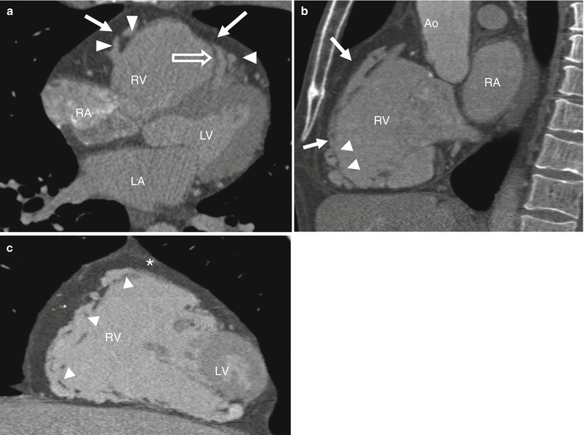
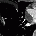
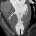
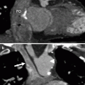
Fig. 7.1
Panel (a): an axial computed tomographic (CT) image in an arrhythmogenic right ventricular dysplasia (ARVD) patient demonstrating profound right ventricle dilatation, extensive low-attenuation fatty tissue in the wall of the right ventricle (white arrows), and prominent trabeculae (white arrowheads). Fatty tissue is also seen in the moderator band (open arrow). Panels (b) and (c) are sagittal and coronal reformations, respectively, and show fat in the RV wall (arrows). Scallops are also noted along the RV wall (white arrow heads). The right ventricle is markedly dilated and the inferior wall is thinned. Also note abundant epicardial fat (asterisks in panel c). RV right ventricle, LV left ventricle, RA right atrium, LA left atrium, Ao aorta
Fatty tissue is identified by CT as low-attenuation tissue (negative Hounsfield units) that does not enhance with contrast. In addition, gated CT may identify global and segmental RV dysfunction that is characteristic of ARVD. The ARVD Task Force Criteria report specific MRI-derived values for RV end-diastolic volume index and for RV ejection fraction that may be applied to CT [2].
7.2 Differential Diagnosis of Myocardial Fat
Myocardial fat is seen in adults with normal hearts and in steatosis associated with obesity, lipomatous atrial septal hypertrophy, old myocardial infarction (MI), cardiac lipoma, and other uncommon conditions such as tuberous sclerosis [4] and muscular dystrophies [5] (Table 7.1).
Table 7.1
Differential diagnosis of fatty infiltration of the myocardium
Type of fat | Segmental location | Intramyocardial location | Myocardial thickness | Ventricle (size/function) |
|---|---|---|---|---|
Physiologic | Anterolateral RV | Full thickness | Normal or increased | Normal/normal (RV) |
Free wall | ||||
RVOT | ||||
Chronic infarct | Coronary artery territory | Subendocardial | Normal or decreased | Normal or dilated/normal or depressed (LV) |
Typically LV | ||||
ARVD | RV and RVOT | Subepicardial | Decreased | Dilated/depressed |
Free wall | ||||
Trabeculae |