AC Joint Separation
Abdul O. Nasiru
Daniel B. Nissman
CLINICAL HISTORY
47-year-old male with a history of shoulder pain secondary to a motor vehicle collision.
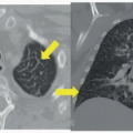
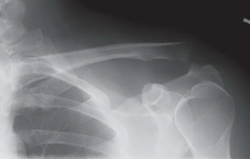 FIGURE 76C |
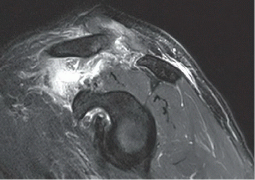 FIGURE 76A |
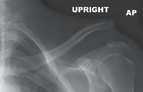 FIGURE 76B |
FINDINGS
AP view of the left shoulder (Fig. 76A) demonstrates inferior subluxation of the acromion at the acromioclavicular joint of one full clavicular shaft width. The coracoacromial distance is increased. Sagittal T2-weighted fat-suppressed MR image (Fig. 76B) demonstrates a discontinuous coracoclavicular ligament complex with a large amount of intervening fluid. AP view of the left shoulder in another patient (Fig. 76C) demonstrates marked relative inferior dislocation of the acromioclavicular joint associated with marked superior displacement of the distal clavicle, resulting in focal convexity of the subcutaneous tissues.
DIFFERENTIAL DIAGNOSIS
Acromioclavicular joint separation with or without coracoclavicular ligament disruption, distal clavicle fracture.
DIAGNOSIS
Related posts:


Stay updated, free articles. Join our Telegram channel

Full access? Get Clinical Tree


