CHAPTER 21 Acquired Cholesteatoma
Epidemiology
Most acquired cholesteatomas result from complications of chronic middle ear infections. Loss of collagen fibers and structural support of the tympanic membrane along with negative middle ear pressure results in a retraction pocket lined with squamous epithelium. Continued negative middle ear pressure and accumulation of epithelial cells, keratin, and cellular decay expand the pocket. Bone destruction of the tympanum and ossicles ensues secondary to direct pressure from the expanding cholesteatoma, biochemical factors related to chronic inflammation and the cholesteatoma itself, and osteoclastic activity. Most cholesteatomas involve the weaker pars flaccida with less common involvement of the pars tensa.
Other potential etiologies of cholesteatoma formation include epithelial invasion through a tympanic membrane perforation, squamous metaplasia of middle ear epithelium, and basal cell hyperplasia.
Clinical Features
Otorrhea and a conductive hearing loss are the most common complaints associated with a cholesteatoma. Patients often have a history of multiple earaches in childhood followed by chronic ear problems. Physical exam demonstrates retraction of the eardrum, often with a perforation, and surroundingbony erosion. Patients with ossicular erosion typically present with a 30- to 60-dB conductive hearing loss. On otoscopic examination, the characteristic clinical finding is the presence of a “pearly white mass” behind the tympanic membrane. However, the tympanic membrane often becomes scarred and retracted in chronic inflammation and the otologist may not be able to accurately assess the middle ear cavity.
Pathology
Cholesteatomas are lined by stratified squamous epithelium and contain desquamated keratin and purulent material.
Treatment
Surgical excision or exteriorization is the treatment of choice. In patients who cannot tolerate surgery, disease can be controlled with repeated cleansing under a surgical microscope.
Imaging Findings
CT
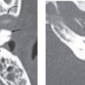
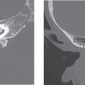
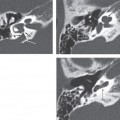
Computed tomography (CT) is the preferred modality when imaging cholesteatomas, as the defining feature is bone destruction. Cholesteatomas typically present as a nondependent middle ear soft tissue mass in a characteristic location associated with bone and ossicular erosion.
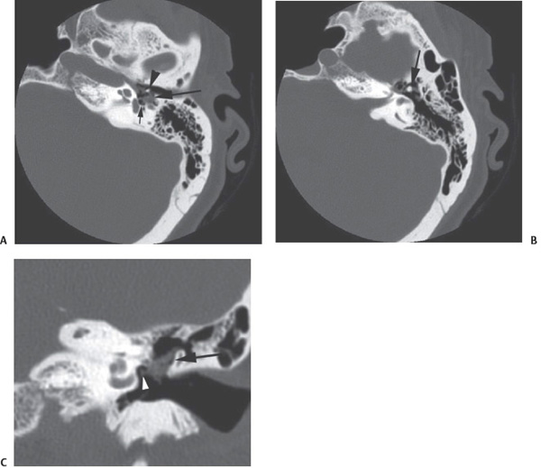
Figure 21-1 Computed tomography (CT) findings of cholesteatoma. (A) Axial image shows a focal soft tissue mass located within the middle ear cavity (large arrow). The manubrium of the malleus (arrowhead
Related posts:

Stay updated, free articles. Join our Telegram channel

Full access? Get Clinical Tree


