KEY FACTS
Terminology
- •
Acute necroinflammatory disease of gallbladder (GB) secondary to stasis and ischemia, not related to gallstones
Imaging
- •
GB wall thickening (> 4 mm) with layered/striated wall
- •
GB distension
- •
Positive sonographic Murphy sign, which may not be elicited if patient obtunded, unconscious, or sedated
- •
US 1st line, HIDA scan for indeterminate US
- •
40% develop complications such as gangrene, perforation, and empyema
- •
CT for complications
Top Differential Diagnoses
- •
Acute calculous cholecystitis
- •
Nonspecific GB wall thickening
- •
GB mucocele
Pathology
- •
Combination of increased bile viscosity and wall Ischemia with secondary infection
- •
Bile cultures positive in up to 78%
- •
Pathogenesis multifactorial: Critical illness with sepsis, shock, recent surgery, trauma, or burns
Clinical Issues
- •
0.2-0.4 % of critically ill patients
- •
Worse prognosis than acute calculous cholecystitis; mortality rate up to 30%
- •
Acute RUQ pain, fever, sepsis in critically ill patient
- •
Nonspecific leucocytosis, elevation of liver function tests
- •
Diagnosis may be challenging in critically ill patient with multiple comorbidities
Scanning Tips
- •
Look for complications such as perforation and abscess
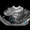
 is shown in a sick patient with a left ventricular assist device. It was not possible to assess the Murphy sign, as the patient was intubated and sedated.
 . There were no gallstones, but there was a small amount of pericholecystic fluid
  .
.
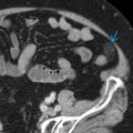
 , wall thickening
  , and a pericholecystic collection
  . No gallstones were found.
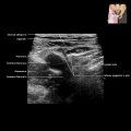
 with intramural edema. There is localized pericholecystic fluid
  with an abscess that is not shown.
Related posts:


Stay updated, free articles. Join our Telegram channel

Full access? Get Clinical Tree


