CHAPTER 29 Adenomatous Lesion
Epidemiology
The neoplasms discussed in this chapter (middle ear adenomas, carcinoids, adenocarcinoma, and endolymphatic sac tumors) are considered different types of adenomatous lesions and are each very rare.
Clinical Features
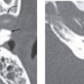
Middle ear adenomas and carcinoids are nonaggressive neoplasms that can present clinically in a manner similar to other more common benign middle ear masses such as primary or secondary cholesteatoma or paraganglioma. They appear as a retrotympanic mass at otoscopy. Symptoms include a feeling of ear “fullness,” tinnitus, and conductive hearing loss. These lesions usually do not involve the facial nerve and therefore do not present with seventh nerve palsy.
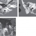
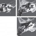
Middle ear endolymphatic sac tumors are more aggressive neoplasms and often present with seventh nerve involvement in addition to the above-described symptoms.
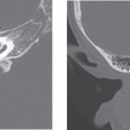
Adenocarcinomas of the middle ear, being an aggressive process, present in a manner similar to other aggressive middle ear processes. Specifically, such lesions present with otorrhea, otalgia, and facial nerve paralysis. These patients often have a long clinical otologic history (e.g., chronic otitis media).
Pathology
Adenomatous (glandular) lesions of the middle ear region include several different entities, all of which are probably histologically related. These include middle ear adenomas, carcinoids, adenocarcinomas, and endolymphatic sac tumors.
Middle ear adenomas and carcinoids most likely represent the same entity classically believed to arise from epithelial tissues of the middle ear. Recent work suggests that they may arise from non-differentiated endodermal stem cells. They are hypovascular lesions demonstrating a nonaggressive, nonpapillary, or pleomorphic histologic pattern with varying amounts of glandular and endocrine differentiation (e.g., carcinoids have more neuroendocrine differentiation than adenomas).
True adenocarcinomas of the temporal bone may arise from the mucosal lining of the middle ear or nondifferentiated endodermal stem cells. Histologically, they have an aggressive, usually nonpapillary appearance. They can invade ossicles or nearby bone and the intracranial compartment.
Endolymphatic duct tumors (also called aggressive papillary adenocarcinomas of the temporal bone) can be seen in the middle ear, though they probably arise in the endolymphatic sac or duct. They are vascular lesions and demonstrate an aggressive papillary histologic pattern.
Treatment
Related posts:

Stay updated, free articles. Join our Telegram channel

Full access? Get Clinical Tree


