GROSS ANATOMY
Overview
- •
Adrenal (suprarenal) glands are part of both endocrine and nervous systems
- •
Lie within perirenal space, bounded by perirenal (Gerota) fascia
- •
Right adrenal is more apical in location
- ○
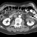
Lies anterolateral to right crus of diaphragm, medial to liver, posterior to inferior vena cava (IVC)
- ○
Often pyramidal in shape, inverted V shape on transverse section
- ○
- •
Left adrenal is more caudal
- ○
Lies medial to upper pole of left kidney, lateral to left crus of diaphragm, posterior to splenic vein and pancreas
- ○
Often crescentic in shape, lambda, tricorn hat, or triangular on transverse section
- ○
- •
Adrenal cortex
- ○
Derived from mesoderm
- ○
Has important endocrine functions
- ○
Secretes corticosteroids (cortisol, aldosterone) and androgens
- ○
- •
Adrenal medulla
- ○
Derived from neural crest
- ○
Part of sympathetic nervous system
- ○
Chromaffin cells secrete catecholamines (mostly epinephrine) into bloodstream
- ○
- •
Adrenal gland has very rich vascular and nervous connections
- •
Arteries
- ○
Superior adrenal arteries : 6-8 from inferior phrenic arteries
- ○
Middle adrenal artery : 1 from abdominal aorta
- ○
Inferior adrenal artery : 1 from renal arteries
- ○
- •
Veins
- ○
Right adrenal vein drains into IVC
- ○
Left adrenal vein drains into left renal vein (usually after joining left inferior phrenic vein)
- ○
ANATOMY IMAGING ISSUES
Imaging Recommendations
- •
Transducer: 2-5 MHz for adults
- •
High-frequency (7.5- to 10.0-MHz) linear transducer used in neonates
- •
Complex shape requires multiplanar evaluation
- ○
Sweeps should be taken in both transverse and longitudinal planes
- ○
- •
Right adrenal gland
- ○
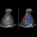
Intercostal transverse approach, using liver as acoustic window
- ○
Direct anterior abdomen scanning is limited by overlying bowel loops and depth to gland
- ○
- •
Left adrenal gland
- ○
Intercostal at midaxillary line, using spleen or left kidney as acoustic window
- ○
In pediatric subjects and thin adults, direct transabdominal US at epigastrium
- –
Stomach may be distended with fluid to serve as acoustic window
- –
- ○
- •
Sonographic appearance
- ○
Relative to body size, adrenal glands are much larger and more easily identified in neonatal population
- –
Also makes them more vulnerable to hemorrhage
- –
- ○
Cortex is hypoechoic with hyperechoic medulla
- –
Creates ice cream sandwich appearance
- –
- ○
Adrenal glands often difficult to see and easily overlooked in adult patients (especially if obese) unless specifically targeted
- ○
Any masses should be documented and measured
- –
Pay particular attention to sonographic appearance (e.g., echogenic mass highly suggestive of fat in a myelolipoma)
- –
- ○
CLINICAL IMPLICATIONS
Clinical Importance
- •
Rich blood supply reflects important endocrine function
- •
Adrenal glands are designed to respond to stress (trauma, sepsis, surgery, etc.) by secreting cortisol and epinephrine
- •
Hemorrhage
- ○
Most common in neonatal population
- –
Associated with many perinatal stressors: Asphyxia, sepsis, birth trauma, coagulopathies
- –
Right > left; bilateral in 5-10%
- –
- ○
Adults
- –
Anticoagulation therapy most common cause
- –
Overwhelming stress (surgery, sepsis, burns, hypotension) may result in adrenal hemorrhage, acute adrenal insufficiency (addisonian crisis)
- □
Relatively uncommon condition but potentially catastrophic event
- □
- –
Blunt abdominal trauma
- □
Generally unilateral (right > left)
- □
- –
- ○
Appearance changes over time
- –
Acute: Hemorrhage appears echogenic & mass-like
- –
Subacute: Blood products liquefy & contract, creating mixed echotexture mass
- –
Chronic: Adrenal resumes normal size, ± Ca²⁺ or cyst
- –
- ○
- •
Metastases : Common site for hematologic metastases (lung, breast, melanoma, etc.)
- •
Adrenal adenoma
- ○
Very common (at least 2% of general population), but most are small, nonfunctioning
- ○
Functional adenomas
- –
Cushing syndrome (excess cortisol) : Truncal obesity, hirsutism, hypertension, abdominal striae
- –
Conn syndrome (excess aldosterone) : Hypertension, hypokalemic alkalosis
- –
- ○
- •
Myelolipoma
- ○
Benign tumor composed of mature adipose tissue and variable amount of hematopoietic elements
- ○
Characteristic echogenic appearance on US
- ○
- •
Pheochromocytoma
- ○
Catecholamine-secreting tumor from adrenal medulla
- ○
Location: Adrenal gland (90%), sympathetic chain from neck to bladder (10%)
- ○
- •
Neuroblastoma
- ○
Most common extracranial solid malignancy in children (median age at diagnosis: 15-17 months)
- ○
Location: Adrenal gland > retroperitoneum > posterior mediastinum
- ○
ADRENAL GLANDS


