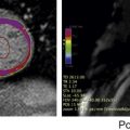
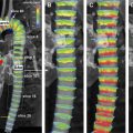
Many novel cardiac MR sequences can be used for assessment of adult patients with congenital heart disease. Although most of these techniques are still primarily used in the research arena, there are many potential applications in clinical practice. Advanced cardiac MR assessment of myocardial tissue characterization, flow hemodynamics, and myocardial strain are promising tools for diagnostic and prognostic assessment late after repair of congenital heart diseases. Novel MR imaging techniques for the assessment of patients with congenital heart disease include 4-dimensional flow imaging, myocardial strain imaging, tissue characterization tools, and 3-dimensional modeling. Current clinical research publications highlight potential applications for these techniques in the near future. Cardiac MR imaging is a well-established method for the diagnosis and follow-up of adult patients with congenital heart disease (CHD). One of the primary roles of MR imaging in these patients is in the postoperative assessment and guidance for the timing of reintervention. Recent advances in cardiac MR imaging have the potential to increase the scope of clinical applications in these patients, expanding its role for not only diagnosis and follow-up but also risk stratification. In this review, recent work on novel cardiac MR imaging methods in adult patients with CHD is emphasized. Velocity-encoded cine MR imaging is a robust technique used clinically for the quantification of blood flow. In patients with CHD, the main application of this technique is to quantify regurgitant volumes and estimate the severity of cardiac shunts. The development of 4-dimensional (4D) flow imaging has many potential applications in adult patients with CHD. The technique consists of a comprehensive evaluation of vascular hemodynamics, achieved through the combined data acquisition of 3 spatial dimensions and 3 blood flow velocity directions during the cardiac cycle. Recent work from Hope and colleagues focused on the application of 4D flow imaging in patients with coarctation of the aorta and bicuspid aortic valve. The authors showed that the quantification of collateral flow in patients with hemodynamically significant coarctation can be accurately obtained with 4D-flow imaging, using 2-dimensional (2D) flow techniques as the standard of reference. In addition to providing comparable collateral flow quantification to 2D flow technique, the 4D flow approach allows for a more comprehensive evaluation of the thoracic aorta flow, including assessment of aortic valve dynamics and estimation of associated aortic valve stenosis owing to bicuspid aortic valve. Finally, the 3-dimensional (3D) acquisition allows for repositioning of the flow measurement planes during the postprocessing analysis, unlike the standard 2D flow technique. Additional applications of 4D flow in patients with CHD have been investigated. A general advantage of the method is to allow for quantification of blood flow in multiple vessels with single acquisition. Therefore, patients with complex CHD can have a mapping of the venous and arterial blood flow obtained in a timely manner. Meadows and colleagues successfully used the single-acquisition 4D approach to evaluate differential pulmonary flow, pulmonary regurgitation, and pulmonary stenosis in patients with tetralogy of Fallot (TOF). Four-dimensional technique permits evaluation of multiple vessels with a single acquisition and allows for the alteration of planes of choice during the postprocessing phase, unlike with 2D flow technique. These characteristics suggest added value for 4D flow imaging in clinical evaluation of patients with CHD. The main limitations for widespread clinical applications of 4D flow include time-consuming data collection and analysis and limited spatial resolution for flow analysis in smaller vessels. The development of optimized fast sequences using sparse sampling techniques shows promise in decreasing scan time and improving spatial resolution. In current clinical practice, steady-state free precession (SSFP) cine images are the most useful sequences for qualitative and quantitative assessment of ventricular function. Volumetric analysis of SSFP images, usually obtained in the short-axis plane, permit quantification of ventricular volumes and ejection fractions. Furthermore, regional function can be quantified by measuring ventricular wall thickening during the cardiac cycle. Advanced techniques for quantification of regional myocardial strain have the potential to identify myocardial dysfunction before depression of global measures of ventricular contractility, such as ejection fraction. Different methods have been proposed for quantification of myocardial strain, which is usually measured in 3 components: circumferential, longitudinal, and radial strain. Applications of myocardial strain imaging in patients with CHD have been a recent focus of clinical research. Ordovas and colleagues described how MR imaging tagging ( Fig. 1 ) can aid in the early detection of left ventricular dysfunction in patients with pulmonary regurgitation after repair of TOF and normal left ventricular ejection fraction and pulmonary regurgitation after repair of TOF. The authors showed that patients with TOF have significantly decreased left ventricular peak circumferential strain in the base and apex levels compared with normal volunteers. Moreover, the left ventricular peak rotation at the basilar and midventricular levels was also delayed in these patients compared with volunteers. Additionally, the same investigators used SSFP cine sequences for quantification of ventricular septal excursion in patients after repair of TOF ( Fig. 2 ). Abnormal ventricular septal excursion was associated with reduced global and septal left ventricular systolic function. It also corresponded with the presence of fibrosis at the left ventricular septum and at the right and left ventricular hinge points, suggesting the technique may be able to identify adverse interventricular interaction.
Key points
Introduction
New clinical applications
Flow Imaging
Quantification of Ventricular Mechanics