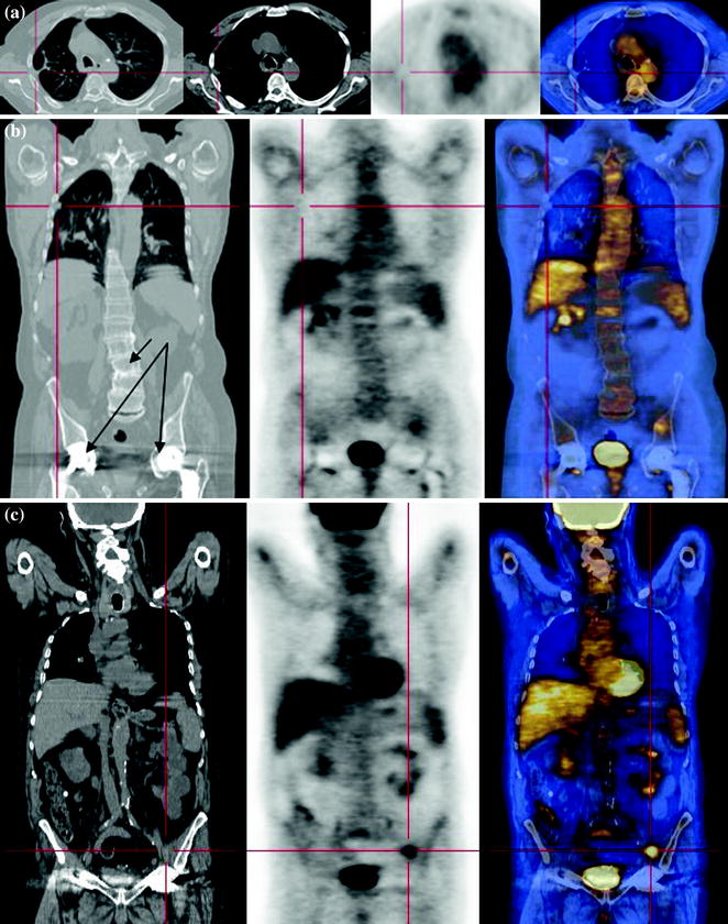
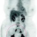
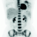
Fig. 61.1
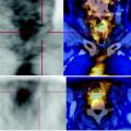
CT-PET shows a solid, patchy nodule with irregular margins, contained within a marked thickening of the left iliac muscle, showing focal glucose consumption. There is no locoregional bone infiltration. At CT, the right iliac wing has osteosclerotic alterations that do not have typical characteristics of a recurrent injury, the absent carbohydrate consumption supporting the hypothesis of a dystrophic condition, or involution, in a patient with bilateral hip prostheses
61.4 Conclusions
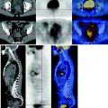
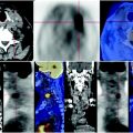
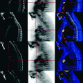
The PET scan shows incomplete response of muscular metastases to chemotherapy. See Fig. 61.2.
