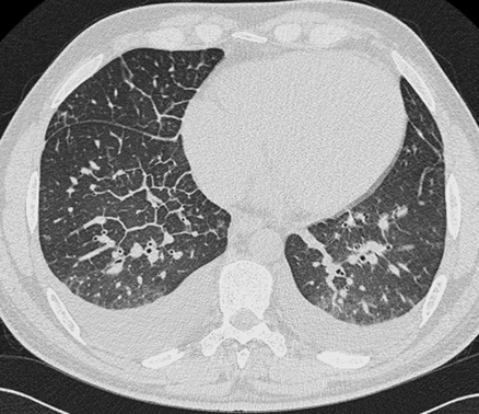
• An excess of extravascular lung water • Interstitial oedema: oedema fluid collecting in a subpleural space manifests as thickening of the interlobar fissures or as a costophrenic recess lamellar ‘effusion’ • Alveolar oedema: this generally spares the apices and extreme lung bases
Airspace disease
AIRSPACE DISEASE
PULMONARY OEDEMA
DEFINITION
 Cardiogenic oedema: increased hydrostatic pressure moves fluid out of the vascular compartment
  this is commonly caused by left heart failure
  it is rarely caused by a reduction in plasma osmotic pressure (e.g. hypoalbuminaemia)
 Non-cardiogenic oedema: this is caused by an increased alveolar-capillary barrier permeability
RADIOLOGICAL FEATURES
CXR/CT
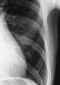
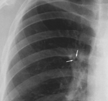
 Kerley B lines: thickened interlobular septa (1–2mm wide, 30–60mm long)
  this occurs within the sub-pleural lung and perpendicular to the pleural surface
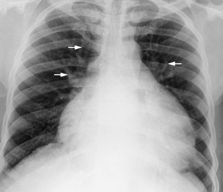
 Peribronchial cuffing: thickened and indistinct airway walls
Cardiogenic oedema
Non-cardiogenic oedema
Distribution
Central ‘bat’s wing’
Tends to be more peripheral
Septal lines
Common
Less common
Peribronchial cuffing
Common
Less common
Pleural effusions
Common
Less common
Cardiomegaly
Yes
No
Pulmonary vasculature
Upper lobe diversion
No redistribution
 Perihilar haze: loss of conspicuity of the central pulmonary vessels
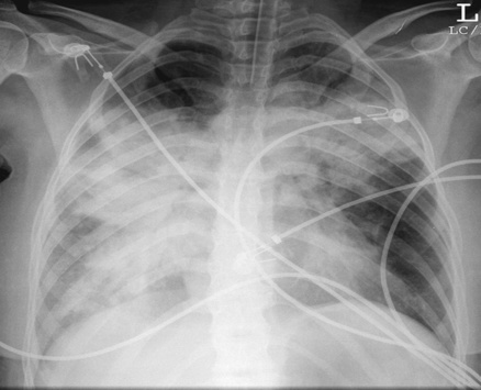
 usually there is bilateral opacification (it can be unilateral)
  opacities may coalesce to produce a general ‘white-out’ (± air bronchograms)
  resolution of any airspace opacification may be rapid (over hours)
 ‘Butterfly’ or ‘bat’s wing’ distribution: this occurs if the central lungs are predominantly affected
Airspace disease
 drowning
  drug induced
  ARDS
ARDS  high altitude
  rapid re-expansion of a collapsed lung
  intracranial disease
 they cross the inner ⅔ of the lung (and tend to point medially towards the hilum)


 the distribution can be affected by coexisting disease (e.g. emphysema can lead to patchy oedema)
 the severity can vary from small symptomless bleeds to life-threatening episodes