Alimentary Tract Vasculature


Alimentary Tract Vasculature


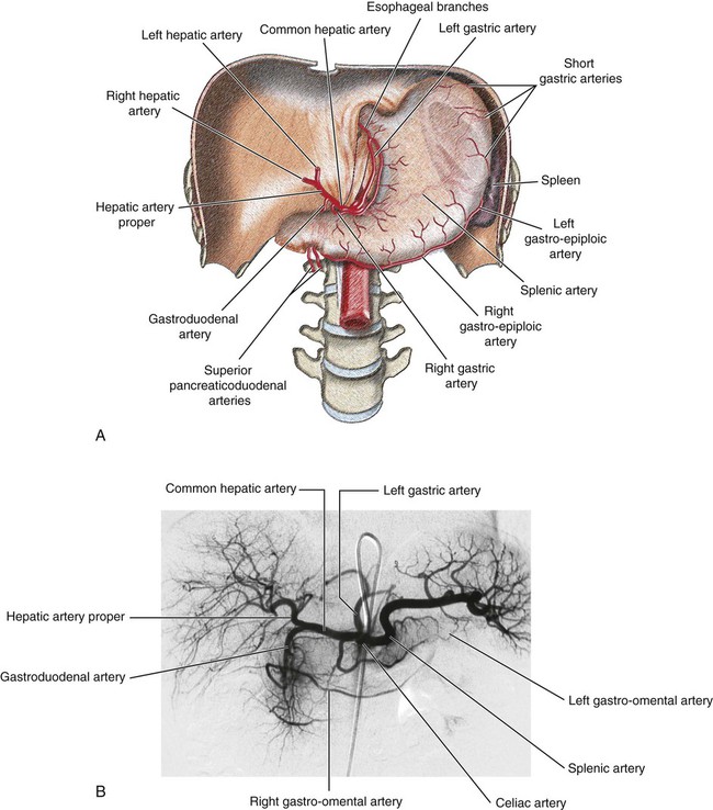
Nadia J. Khati, Morvarid Alaghmand, Michael J. Manzano and Anthony C. Venbrux


Clinical Relevance


An understanding of the vascular anatomy of the alimentary tract allows a wide range of therapeutic options for patients who traditionally were treated by open surgery. In addition to the use of catheter-directed embolotherapy and pharmacologic vasoconstriction for gastrointestinal (GI) hemorrhage, a knowledge of variant anatomy may prevent complications such as nontarget organ embolization. For example, recognition of variant anatomy during hepatic artery chemoembolization for liver tumors may prevent inadvertent delivery of toxic chemotherapeutic drugs to the upper GI tract (e.g., inadvertent reflux of chemotherapeutic agents into the gastroduodenal artery, with possible resultant duodenal mucosal injury and ulceration).


The venous anatomy in the alimentary tract is also important. The transjugular intrahepatic portosystemic shunt (TIPS) procedure functionally creates a portosystemic shunt that reduces the high pressure in patients with advanced portal hypertension. Such elevated pressure may cause life-threatening esophageal variceal hemorrhage. TIPS, though not perfect, can be lifesaving.


Delayed strictures of the biliary system may occur if the hepatic artery is injured (e.g., clipped) during cholecystectomy in a patient with advanced liver disease. Such ischemic strictures result from inadequate blood flow to the bile ducts, the latter receiving blood supply from the hepatic artery.


Thus, an understanding of alimentary tract vasculature is essential while performing image-guided interventions.


Vascular Imaging of the Alimentary Tract


Evaluation of the gastrointestinal tract vasculature can be performed with different imaging modalities including multidetector-row computed tomography (MDCT), magnetic resonance angiography (MRA), conventional angiography and ultrasound.


The most common indications for imaging of the gastrointestinal (GI) tract vasculature include acute and chronic mesenteric ischemia, median arcuate ligament syndrome, aneurysms and dissections as well as evaluation of the portal venous system either in the assessment of portal hypertension or following a transjugular intrahepatic portosystemic shunt (TIPS) procedure.


Ultrasound


The use of abdominal ultrasound in the evaluation of the gastrointestinal (GI) tract vasculature is limited to visualization of the larger vessels such as the major branches of the aorta (celiac trunk, superior mesenteric artery) and the portal venous system. The many branches of the small bowel mesentery and inferior mesenteric artery are usually not visualized due to their small. Other limitations of US include patient body habitus, sonographer’s degree of experience and overlying bowel gas. The 2 most common clinical situations where US is helpful in making a diagnosis is in the evaluation of chronic mesenteric ischemia due to median arcuate ligament syndrome and when evaluating the portal venous system. Using color and Doppler sonography, both entities can easily be diagnosed by measuring velocities across vessels, direction of flow and patency of vessels.


CT Angiography and MR Angiography


CT angiography (CTA) and MR angiography (MRA) are the 2 most commonly used non-invasive imaging modalities for the diagnosis of mesenteric ischemia in the acute setting. Both exams involve the administration of iodinated contrast for CT and gadolinium for MRI and can only be performed in patients with normal renal function. With the advent of volumetric acquisition of images and three-dimensional (3D) reconstruction algorithms both the mesenteric arterial and venous system can be adequately visualized, especially when using dual-phase imaging (arterial and venous phases). Both CTA and MRA are highly accurate in the diagnosis of acute mesenteric artery thrombosis, mesenteric and portal venous thrombosis by demonstrating either a filling defect within the occluded vessels or an abrupt cut-off of the affected vessels. In addition, these modalities may show secondary signs of ischemia such as bowel wall thickening and mesenteric edema or hemorrhage. In patients with renal impairment, non-contrast enhanced MR angiography using balanced steady-state free precession (SSFP) can be used as an alternative to image the aorta and its branches. Some advantages of using CTA over MRA include greater availability in most institutions, faster acquisition time, and improved spatial resolution allowing better visualization of smaller peripheral vessels of the mesenteric circulation. Furthermore, CTA can evaluate the presence of atherosclerotic disease by visualizing calcified plaque, which is very difficult to perceive on MRA.


Catheter Angiography


Catheter angiography is still considered by many radiologists as the gold standard for imaging the aorta and the mesenteric arteries and veins even though it remains an invasive test, involves the use of ionizing radiation and the administration of iodinated contrast material. It remains the study of choice when non-occlusive mesenteric ischemia is suspected. In patients with renal impairment, carbon dioxide (CO2) can be safely substituted for iodinated material as the contrast medium. One of the biggest advantages of using conventional angiography over other imaging modalities is that it can serve as a diagnostic and therapeutic tool when necessary. Vasodilators or thrombolytics can be administered at the time of the examination in addition to visualizing possible collateral circulations.


Arterial Supply


Three major arteries that supply the alimentary tract are the celiac axis, superior mesenteric artery (SMA), and inferior mesenteric artery (IMA).


Celiac Trunk


The celiac axis arises from the ventral surface of the aorta at the level of the T12-L1 disc space. It immediately divides into three branches—the left gastric artery, splenic artery, and common hepatic artery—and supplies the liver, spleen, stomach, and pancreas (Fig. 52-1). Both the splenic and left gastric artery can arise directly from the aorta as separate branches, but in over 95% of patients, the common hepatic artery arises from the celiac trunk.


The left gastric artery is the smallest branch of the celiac axis. It supplies the gastroesophageal junction (abdominal part of the esophagus) and anastomoses with esophageal branches from the thoracic aorta. It also supplies the fundus and a portion of the body of the stomach and anastomoses with the right gastric artery, a branch of the hepatic artery.


The splenic artery is the largest branch of the celiac trunk. It has a tortuous course to the left along the superior border of the pancreas and has three branches:


The common hepatic artery is a medium-sized branch of the celiac trunk and runs to the right. It divides into two major branches:


Other branches of the common hepatic artery include the supraduodenal and right gastric arteries.


Anastomoses in the stomach include:


Dec 23, 2015 | Posted by in INTERVENTIONAL RADIOLOGY | Comments Off on Alimentary Tract Vasculature

Full access? Get Clinical Tree

Get Clinical Tree app for offline access