Aortic Stent-Grafts


Aortic Stent-Grafts


Raghuveer Vallabhaneni and Mark A. Farber


Clinical Relevance


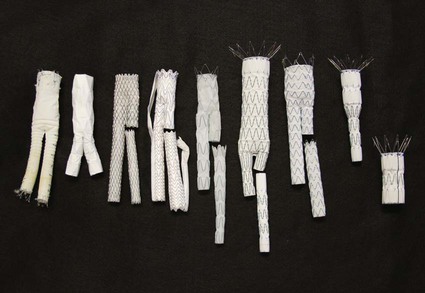
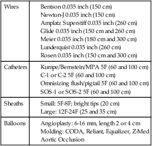
Over the past decade, stent-graft use for the treatment of aortic pathologies has steadily increased. Over 64% of infrarenal aortic aneurysms were treated with endovascular aneurysm repair (EVAR) in 2007.1 Expanded indications have occurred as additional experience has been obtained using this technology and includes use of stent-grafts to treat aneurysms in the pararenal and visceral aorta, as well as emergent and nonaneurysmal pathologies.2,3 This, however, has been associated with a rise in complication rates as devices are being implanted outside their intended “instructions for use” (IFU) guidelines.46 To meet this demand for greater application of endovascular techniques, medical device manufacturers have improved stent-graft design (Fig. 11-1) with new design modifications that are currently being evaluated in clinical trials. Hopefully, such trials will provide data to enable the clinician to apply newer devices to increasingly complex aortic pathologies that have traditionally been treated by open procedures.7


Indications


Infrarenal Aneurysms


Current indications for stent-graft implantation in the infrarenal aorta involve the treatment of abdominal aortic aneurysms (AAAs). Strict indications include the presence of an AAA with a greater than 5 cm diameter, rapidly expanding aneurysms (>0.5 cm in 6 months), or aneurysms greater than two times the normal aortic diameter. The patient should also be a suitable candidate for endovascular exclusion based on anatomic criteria. Although these specifications differ slightly based upon which device is being implanted, they generally include the following characteristics. For an infrarenal aortic stent-graft, the aortic neck must be of suitable diameter and quality to provide an adequate seal. This is considered to be at least 10 mm in length, relatively free of significant calcification and thrombus, a nonaneurysmal neck (18-32 mm in diameter) with parallel walls, and angled less than 60 degrees. The anatomic limitations for the access vessels are less strict. Iliac stenosis, calcification, and tortuosity should allow for passage of the devices and have adequate length for sealing regions. Preservation of one hypogastric artery for pelvic blood flow is suggested, but isolated cases of bilateral internal iliac artery exclusion have been reported.8,9 New iliac-branched devices are in clinical trials in the United States and may help preserve hypogastric flow in patients who do not have an adequate sealing zone in the common iliac arteries.10


Pararenal Aneurysms


Aortic aneurysms in the region of the renal arteries are grouped collectively as pararenal aneurysms and further classified into juxtarenal and suprarenal subtypes. There is no universally agreed-upon definition of the term juxtarenal aneurysm, but it is commonly used to describe a complex AAA with either a short infrarenal neck or one that encroaches upon the renal segment of the aorta. Suprarenal aneurysms involve renal arteries and extend up to the splanchnic arteries. Type IV thoracoabdominal aortic aneurysms extend to a variable abdominal length but always involve the visceral aortic segment. Classification systems have been proposed, but none have gained wide acceptance or clinical use.11 This lends some ambiguity to the terms and makes comparison of clinical studies difficult.


Until recently in the United States, the only way to treat pararenal aneurysms using endovascular techniques was to modify grafts to accommodate the anatomy of the individual patient or to use “off-label” current devices and technology. These techniques include handmade placements of fenestrations or scallops, openings in the graft material of the stent-graft to accommodate the visceral vessels, or use of a “snorkel” or “chimney” technique where stents are placed by upper extremity access through visceral vessels and extended superior to the graft material of an EVAR device to allow blood to flow to the visceral vessels. As of the writing of this chapter, there are two U.S. Food and Drug Administration (FDA)-approved clinical trials currently underway for fenestrated devices for treatment of juxtarenal aneurysms. The first trial, which is nearing completion (phase III), is the Cook Zenith fenestrated device (Cook Medical, Bloomington, Ind.). This device is indicated for use in aneurysms with a 4-12 mm neck and less than 45 degree of angulation at the neck.7 The second trial, beginning enrollment shortly, is from Endologix (Irvine, Calif.) for an “off-the-shelf” fenestrated device.


Also under investigation are branched devices. In these devices, a branch graft is sewn onto the device, the visceral branch is cannulated through the branch graft, and a stent is deployed into the vessel from the branch for support. However, no trials for such devices are yet underway in the United States.


Other aortic pathologies that have been treated with endovascular devices include ruptured AAA, penetrating atherosclerotic ulcers, mycotic aneurysms, aortoenteric fistulas, emboligenic lesions, and traumatic aortic injuries.2,12,13 Although not designed for these entities, many times the devices function extremely well, since there is an increased surface area contact with the device, thereby limiting device migration issues. However, limitations do exist based upon appropriate sizing and small aortic bifurcations. Long-term data regarding the treatment of infected pathologies is limited. Use of endovascular devices as a temporizing method in these emergent high-risk operations is appealing.


Contraindications


Successful EVAR is directly related to appropriate patient selection. Preprocedural computed tomographic angiography (CTA) with axial and multiplanar reformatted imaging can adequately identify and delineate the patient’s anatomic characteristics. Axial imaging alone has some shortcomings with respect to accurately sizing tortuous anatomy.14


Absolute contraindications to infrarenal EVAR include those patients whose aortic neck is too short or too large to allow for exclusion of the aneurysm. EVAR would be contraindicated if repair requires occlusion of a critical branch vessel. This may include a patient with a patent inferior mesenteric artery when preexisting superior mesenteric artery and celiac artery stenosis or occlusion is present, accessory renal arteries supplying significant renal parenchymal mass (e.g., horseshoe kidney), and rarely a dominant lumbar artery providing critical blood supply to the spinal cord. Significant thrombus in the aortic landing zone may also be a relative contraindication for fear of embolization or lack of adequate seal. Additionally, alternative therapies or devices should be sought in patients with allergies to material components in a device’s composition.


Relative contraindications included patients whose anatomic criteria do not completely fit IFU guidelines. This may include patients with small or diseased iliac arteries that require a conduit for device implantation. Finally, in obese patients for whom adequate imaging either during the implantation procedure or during follow-up is required, careful consideration of both the short- and long-term patient risks is necessary.


In patients who have pararenal or type IV thoracoabdominal aneurysms and are not candidates for open repair, fenestrated endovascular aortic repair (FEVAR) or branched endovascular aortic repair (BEVAR) may be options. However, clinical trials must be completed prior to these devices being commercially available. Physician-modified grafts may be considered in these patients, although they would be performed outside the devices’ IFU guidelines.


Equipment


The equipment needed to perform EVAR involves typical interventional sheaths, wires, and catheters, with the addition of larger sheaths and longer and stiffer wires. Table e11-1 details appropriate equipment, but individual preferences can be substituted in many instances. Initial access wires and sheaths should include starter wires and 5F to 8F sheaths. Three grades of wire stiffness can be used, and in order of increasing stiffness include the Amplatz Superstiff, Meier, and Lunderquist wires. Each of these wires has its own inherent characteristics and can be applicable in different situations, depending on the interventionalist’s preferences and patient’s anatomy. Although some devices can be inserted over 180-cm-long wires, 260-cm or longer lengths are typically needed. Larger sheaths are sometimes necessary to help accommodate iliac tortuosity and should include sizes from 16F through 24F. Now that most devices, except the Gore Excluder and TAG (W.L. Gore & Associates, Flagstaff, Ariz.), are designed for sheathless insertion, sheaths are becoming less necessary. The presence of significant iliac tortuosity may require utilization of rigid sheaths to allow completion of the procedure. Occasionally, sequential dilators are necessary to determine whether iliac access is possible prior to opening a device.


Additional ancillary equipment includes an adequate imaging system (12-inch image intensifier minimal), power injector, intravascular ultrasound, occlusion balloons, large Palmaz stents, and molding balloons. In some institutions where percutaneous access is preferred, appropriately sized closure devices should be available. As lower-profile devices become available, this may be a more appealing option. Appropriate interventional equipment should also be available to handle potential complications involving the great vessels, renal arteries, and access vessel complications.


Technique


Anatomy and Approach


The approach involves either percutaneous or open surgical exposure of the common femoral arteries. Oblique incisions are generally preferred unless femoral reconstructions are planned for preexisting occlusive disease. Care should be taken during the dissection not to transect the lymphatics. Ligation or clipping of any visible lymphatic vessels should be performed. Once the femoral sheath is entered, the artery is identified as it exits under the inguinal ligament, and it is encircled with Silastic or cloth tapes both proximally and distally. Systemic heparinization is instituted at this juncture with administration of 80 to 100 units/kg heparin intravenously. Once this has been completed, bilateral aortic access is then obtained using standard interventional techniques. Catheter exchange can then occur so that a stiff wire (Amplatz, Meier, or Lunderquist) is placed through the ipsilateral femoral artery, and a diagnostic catheter on the contralateral side. General interventional principles should be followed, especially with respect to monitoring the end of the stiff wires, as well as fluoroscopic imaging during the insertion of the device.


After the primary device is prepped and initial orientation determined, the main device is inserted through the ipsilateral iliac artery and positioned near the renal arteries. Based on device selection, it is typically deployed just below the renal arteries with fluoroscopic and injected-contrast guidance. Implantation of the device too far below the renal arteries has been associated with device migration and a higher failure rate. If the system being implanted is a modular component system, iliac extensions are inserted and deployed. These iliac limbs are typically extended to the level of the common iliac bifurcation bilaterally, facilitating both stabilization of the device and exclusion of the aneurysm by preventing retrograde flow. After device implantation, balloon “molding” is often performed. This process confirms expansion of the device to full conformation, facilitates achievement of a satisfactory seal zone between modular components, and may eliminate areas of residual luminal narrowing that predispose to early thrombosis. Exclusion of the aneurysm is then confirmed by angiography. Occasionally, detailed views must be obtained to ensure appropriate device attachment at the level of the renal arteries and in the iliac system. Terminating the procedure with a significant endoleak can be problematic and places the patient at continued risk for aneurysm rupture.


The most important component of successful fenestrated aneurysm repair is careful and accurate advanced planning of the procedure and graft construction. CTA allows measurements of distances using centerline-of-flow analysis, and of the clock position of the target vessel using axial measurements. The criteria for device implantation are essentially unchanged from standard endovascular repairs. The proximal landing zone must consist of at least 2 cm of normal parallel aortic wall, less than 32 mm in diameter for juxtarenal aneurysms, and less than 38 mm for thoracoabdominal aneurysms. The centerline measurement from the top of the landing zone to the center of the target vessel origin is recorded, as are the clock position, orientation, and diameter of each target vessel origin. The device configuration can consist of single or multiple fenestrations, depending on patient characteristics. Small fenestrations (6 × 8 mm) are preferentially designed for branched stent-grafts or vessels arising from the aneurysmal aorta. Large fenestrations (8 × 10 mm) are used preferentially for fenestrated stent-grafts or vessels arising in the proximal seal zone of normal aorta.


For placement of the Cook fenestrated device now in trials, the larger iliac system is used for the main body of the device, and a 24F sheath is placed in the contralateral side. The 24F sheath is then punctured with a needle, and multiple 5F sheaths are sequentially placed inside of it. Using angled catheters, the visceral vessels are cannulated with wires to assist in orientation of the fenestrations with the vessels. The brachial artery is also accessed and commonly used to cannulate the superior mesenteric artery. Catheterization of the renal arteries and superior mesenteric artery is verified with selective contrast injections. Once visceral access is confirmed, the main body is visualized under fluoroscopy to confirm orientation. It is then placed through the ipsilateral femoral artery. Through the contralateral sheath, each vessel is then cannulated through the fenestration as the previous marking wire is removed. A balloon-expandable stent-graft is introduced into each target vessel after it has been selectively catheterized through the stent-graft fenestration. Once all bridging stent-grafts are in place, the main-body stent-graft is fully deployed. The balloon-expandable stent-grafts are then deployed to profile and flared proximally with a balloon. A bifurcated modular stent is then deployed below the fenestrated main body into bilateral common iliac arteries.


Technical Aspects


Careful inspection of the renal artery and renal blood flow are critical at the completion of the procedure. If possible, patients with preexisting renal artery stenosis should be stented at the completion of the procedure or during the follow-up period. Placement of renal stents prior to EVAR can cause renal complications and make device implantation more difficult. Maneuvers can help minimize contrast use in patients with renal insufficiency by using intravascular ultrasound to locate the renal arteries and implant the device. However, experience is needed with this technique to ensure proper visualization.


Initially the iliac limbs of devices were only inserted a short distance into the common iliac arteries, with the primary intention of excluding retrograde arterial flow. The length of iliac limb extension was often guided by the distal location of the main-body device. As techniques have evolved, many clinicians now cover the entire length of the common iliac arteries, and additionally make individualized decisions regarding the appropriate length of the main-body device. The optimal location of the distal aspect of the main-body device is based upon the curvature and unique anatomic features of the aneurysm. Consideration of these factors reduces the risk of migration of the device.


When dealing with large sheaths in the iliac arteries, one important consideration is prolonged limb ischemia. These sheaths are commonly occlusive in not only the external iliacs but also the hypogastric arteries. In complex fenestrated or branched endovascular cases, it is important to minimize ischemia by careful planning and proceeding in an efficient manner. If a case is known to be lengthy, it may be worthwhile to place 5F sheaths in the common or superficial femoral arteries in an antegrade fashion. These sheaths can then be connected to the larger sheaths to maintain continuous blood flow and minimize ischemic time to the legs. Temporary axillobifemoral bypass has also been reported as an adjunct measure in these procedures to help minimize leg ischemia.15


When diseased or tortuous iliac arteries are present, additional maneuvers may be necessary to allow insertion of the delivery catheter. This can involve one of several techniques that includes stiffer or secondary “buddy” wire, dilatation or angioplasty of isolated stenosis, retrograde iliac endarterectomy, or placement of an iliac conduit. An endoconduit may also be performed, where a covered stent is placed inside the stenotic artery and ballooned to the appropriate size.16 Recanalization of occluded iliac arteries using subintimal angioplasty has also been reported. Each of these techniques contains individual risks and benefits that must be weighed in each patient.

Only gold members can continue reading. Log In or Register to continue

Stay updated, free articles. Join our Telegram channel

Dec 23, 2015 | Posted by in INTERVENTIONAL RADIOLOGY | Comments Off on Aortic Stent-Grafts

Full access? Get Clinical Tree

Get Clinical Tree app for offline access