KEY FACTS
Terminology
- •
Localized pus collection in liver due to Entamoeba histolytica with destruction of hepatic parenchyma
Imaging
- •
General feature
- ○
Most often solitary (85%), peripherally located
- ○
- •
Ultrasound
- ○
Sharply demarcated, round or ovoid mass
- ○
Hypoechoic with low-level internal echoes
- ○
May see internal septa or wall nodularity
- ○
May see posterior acoustic enhancement
- ○
- •
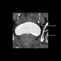
CECT
- ○
Typically hypoattenuating unilocular lesion
- ○
Peripheral rim or capsule enhancement
- ○
May see hypodense halo due to edema
- ○
Top Differential Diagnoses
- •
Hepatic metastasis (post treatment, cystic, or necrotic)
- •
Hepatic pyogenic abscess
- •
Hepatic hydatid cyst
- •
Biliary cystadenoma/cystadenocarcinoma
- •
Infarcted liver after transplantation
Pathology
- •
Entamoeba histolytica
- •
Primary source of infection: Human carriers passing amebic cysts into stool
Clinical Issues
- •
Right upper quadrant pain, tender hepatomegaly, diarrhea with mucus
- •
Indirect hemagglutination positive in 90% of cases
Scanning Tips
- •
Rule out pyogenic or fungal abscess, cystic lesions
- •
Check for history of transplantation, ablation, or chemotherapy for liver tumor or metastasis, which may simulate amebic abscess on imaging
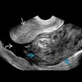
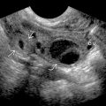
 and central anchovy paste consistency of contents
  .
.
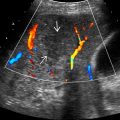
 . The contents are heterogeneous due to floating debris
  . Also note the mild posterior acoustic enhancement
  .
.
 .
.Related posts:

Stay updated, free articles. Join our Telegram channel

Full access? Get Clinical Tree


