Volume of amniotic fluid (AF) represents a balance between
1. Mechanisms producing AF—fetal urine flow, oronasal, and lung secretions
2. Mechanism allowing passage of fluid into the amniotic cavity—fluid movement through the chorion frondosum and membranes, highly permeable fetal skin
3. Mechanism involved in uptake of AF—fetal swallowing
Fetal urination is the major source of AF in the second half of pregnancy
Increase in intramembranous absorption of AF leads to decrease in AFV in cases with chronic severe placental insufficiency
Echogenic AF is the presence of particles in amniotic fluid (represent either meconium or vernix)
Usually not associated with any adverse pregnancy outcome
1. It cushions the fetus against physical trauma.
2. Allows for capacious growth of the fetus without any distortion by adjacent structures.
3. Imparts thermally stable environment.
4. Helps to prevent infection.
5. Source of nutrients to the developing embryo.
Amniotic fluid index technique
Divides the uterus into four quadrants using maternal sagittal midline vertically and transverse line approximately halfway between symphysis pubis and uterine fundus (Figure 22.1).
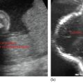
Select the deepest AF pocket free of umbilical cord and fetal parts. The greatest vertical dimension is measured with USG transducer perpendicular to the floor.
(Maximum vertical pocket [MVP]) >2 centimeters—normal
—1–2 centimeters—reduced
<1 centimeter—oligohydramnios
Stay updated, free articles. Join our Telegram channel

Full access? Get Clinical Tree


