Presentation and Presenting Images
A 58-year-old female presents for routine screening mammography.
43.2 Key Images
( ▶ Fig. 43.3, ▶ Fig. 43.4, ▶ Fig. 43.5, ▶ Fig. 43.6)
43.2.1 Breast Tissue Density
The breasts are heterogeneously dense, which may obscure small masses.
43.2.2 Imaging Findings
The imaging of the left breast is normal (not shown). A new group of faint calcifications (circle) is noted in the upper region of the right breast in the posterior depth, 10 cm from the nipple on the mediolateral oblique (MLO) view ( ▶ Fig. 43.3). The calcifications are not well seen on the craniocaudal (CC) view ( ▶ Fig. 43.1). The MLO tomosynthesis movie can be used to localize the calcifications in the CC view. The MLO tomosynthesis movie ( ▶ Fig. 43.4) localizes the group of calcifications to the lateral portion of the breast, placing them at the 10 to 11 o’clock location, 10 cm from the nipple ( ▶ Fig. 43.5 and ▶ Fig. 43.6). This localization is helpful for further diagnostic mammographic evaluation.
43.3 BI-RADS Classification and Action
Category 0: Mammography: Incomplete. Need additional imaging evaluation and/or prior mammograms for comparison.
43.4 Diagnostic Images
( ▶ Fig. 43.7, ▶ Fig. 43.8, ▶ Fig. 43.9)
43.4.1 Breast Tissue Density
The breasts are heterogeneously dense, which may obscure small masses.
43.5 Imaging Findings
Additional magnification imaging ( ▶ Fig. 43.7 and ▶ Fig. 43.8) reveals a group of amorphous calcifications. This new finding has a low suspicion of malignancy and underwent stereotactic biopsy. The radiograph of the specimen ( ▶ Fig. 43.9) from the stereotactic biopsy demonstrates the faint amorphous calcifications (circles).
43.6 BI-RADS Classification and Action
Category 4A: Low suspicion for malignancy
43.7 Differential Diagnosis
Sclerosing adenosis: Stereotactic biopsy ( ▶ Fig. 43.7) was performed following a diagnostic evaluation revealing grouped amorphous calcifications. The pathology was consistent with sclerosing adenosis.
Fibrocystic changes: Fibrocystic changes commonly present as hazy, small, faint calcifications. This diagnosis would be considered concordant if the radiologist felt the calcifications had been adequately sampled.
Ductal carcinoma in situ (DCIS): DCIS may present as amorphous calcifications. This would be a concordant biopsy.
43.8 Essential Facts
With new or increasing calcifications, the next step is diagnostic mammography with magnification views. Magnification views cannot be performed with tomosynthesis.
Tomosynthesis is helpful in localizing findings, both calcifications and masses, seen on one mammographic view. .
Stereotactic biopsy is recommended for new calcifications with suspicious morphology. Calcifications seen best on tomosynthesis may be biopsied using tomosynthesis-directed stereotactic-guided biopsy.
43.9 Management and DBT Principles
Spangler and colleagues (2011) found that tomosynthesis was slightly less sensitive than conventional mammography for the detection of calcifications. The authors felt that with improvements in processing algorithms and display, tomosynthesis could potentially improve in the detection of calcifications.
Kopans and colleagues (2011) found that calcifications can be seen with the same or better clarity on tomosynthesis as on conventional mammography, and tomosynthesis allows for the same and perhaps improved analysis of calcifications.
Magnification views are performed with conventional mammography; magnification views are not possible with tomosynthesis. Tomosynthesis systems must support both conventional mammography and tomosynthesis.
43.10 Further Reading
[1] Berg WA, Arnoldus CL, Teferra E, Bhargavan M. Biopsy of amorphous breast calcifications: pathologic outcome and yield at stereotactic biopsy. Radiology. 2001; 221(2): 495‐503 PubMed
[2] Kopans D, Gavenonis S, Halpern E, Moore R. Calcifications in the breast and digital breast tomosynthesis. Breast J. 2011; 17(6): 638‐644 PubMed
[3] Spangler ML, Zuley ML, Sumkin JH, et al. Detection and classification of calcifications on digital breast tomosynthesis and 2D digital mammography: a comparison. AJR Am J Roentgenol. 2011; 196(2): 320‐324 PubMed

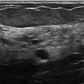
Fig. 43.1 Right craniocaudal (RCC) mammogram.
Related posts:


Stay updated, free articles. Join our Telegram channel

Full access? Get Clinical Tree


