KEY FACTS
Imaging
- •
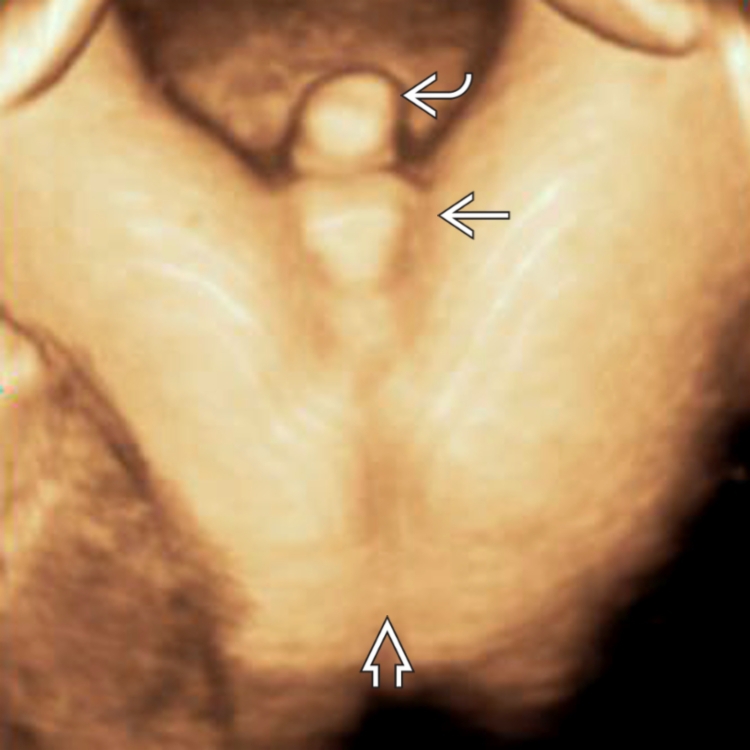
Inability to demonstrate normal anal dimple or target sign
- •
Normal appearance
- ○
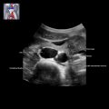
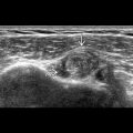
Axial: Hyperechoic mucosa within hypoechoic muscular ring
- ○
Coronal: Hyperechoic mucosal stripe between hypoechoic walls, extending to perineum
- ○
No other appearance is proof of normal anorectal development; should be demonstrable from 20 weeks
- ○
- •
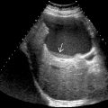
Dilated, fluid-filled distal bowel is late manifestation; not seen at time of 2nd-trimester survey
- ○
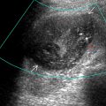
U- or V-shaped bowel in presacral space without extension to perineum
- ○
- •
Polyhydramnios unusual
- ○
Unlike proximal small bowel atresias
- ○
Scanning Tips
- •
Remember that colon is often prominent in 3rd trimester
- ○
Normal caliber ≤ 18 mm
- ○
- •
3D data sets can be obtained to demonstrate rectum in sagittal and coronal planes
- •
Look for calcified meconium enteroliths
- ○
Results from mixture of meconium and fetal urine within bowel lumen if fistula between colon and bladder
- ○
Look for echogenic “marbles” moving within bowel
- ○
- •
Look for other abnormalities as anal atresia is seldom isolated
- ○
VACTERL association ( v ertebral, a nal atresia, cardiac, t racheo- es ophageal, r enal and l imb anomalies)
- ○
Cloacal exstrophy
- –
Abdominal wall defect, absent bladder, often spine abnormalities
- –
- ○
Cloacal malformation
- –
Female fetus, distended vagina, small bladder
- –
- ○
- •
Take dedicated views of rectum/anus when any bowel, spine, or genital abnormality is present
 but no evidence of an anal dimple in the expected location. A normal anal dimple (target sign)
  is shown in the inset.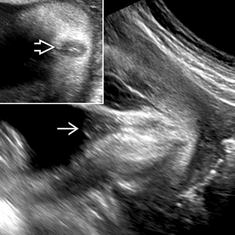
 (empty scrotum); short, curved penis
  ; and absence of an anal opening in the natal cleft
  . Anal atresia is often associated with genital and distal spine abnormalities. Look for the anal dimple when evaluating fetal sex.