CHAPTER 3 ![]()
Anatomy, Physiology, and Pathology of the Breast
Summary of Important Points
MAMMARY GLANDS
• Mammary glands are accessory glands of the female reproductive system. They are a specific type of sudoriferous or apocrine sweat glands specialized in manufacturing colostrums and milk, which is needed for the newborn. Mammary glands are also sometimes referred to as modified sebaceous glands.
• In humans the mammary glands are also regarded as a sexual asset therefore the loss or scarring of these glands can have devastating psychological implication for women.
Shape and Size of the Breast
• Spherical in shape with size varying with age, menstrual cycle, and lactation.
• Hormonal stimulation causes the breasts to grow, and will also affect breast shape and size.
Muscles Associated with the Breast
• The pectoralis major muscle lies immediately below the breast.
• Serratus anterior lies to the lateral aspect of each breast.
• The pectoralis minor muscle, a thin flat triangular muscle, lies deep to the pectoralis major muscle and covers the serratus anterior and the external oblique muscles.
• The latissimus dorsi muscle runs from the lateral aspect of the breast across the midaxillary line to the lower back.
Retromammary Space
• The retromammary space is a layer of adipose or fatty tissue that separates the breast from the pectoral muscle.
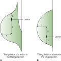
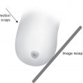
• The retromammary space is seen on the mediolateral projection in mammography imaging but only on 20% of the craniocaudal projections.
Exact Location of the Breast
• The breast extends vertically from the clavicle at the second or third rib, to meet the abdominal wall at the level of the sixth or seventh rib.
• Medially the breast extends from the midsternum to the midaxillary line and to the latissimus dorsi muscle, laterally.
Surface Anatomy of the Breast
• The skin of the breast, like the skin of the body, is filled with sweat glands, sebaceous glands (oil glands), and hair follicles that produce hair on the skin surface. The skin of the breast is thickest at the base, thinning as it approaches the nipple.
• The nipple lies at the center point of the breast. The nipple itself contains multiple crevices, within which are 15-20 orifices, or ducts that transfer milk from the lactiferous ducts to the exterior. Normally the nipple can be either flattened or inverted, both unilaterally and bilaterally. However, a nipple that suddenly becomes inverted or flattened can indicate malignancy.
• The areola is the smooth, circular darkened area surrounding the nipple and contains many small protrusions on its surface called Morgagni tubercles. These tubercles are formed by opening of the ducts of the Montgomery glands, specialized sebaceous-type glands found on the areola. Montgomery glands usually become more prominent during pregnancy and lactation. The glands secrete a fatty lubricant, which protects the nipple during nursing.
• The pigmentation of the areola is partly dependent on estrogen levels. In younger women it is more prominent, but tends to fade at menopause. However, estrogen use at any age will increase pigmentation of the areola.
Location Terminology
• The breast is generally described in terms of the face of a clock or 4 quadrants. The breast can also be divided into 4 regions.
Four-Quadrant Method
• The 4 quadrants are: the upper-outer quadrant (UOQ), upper-inner quadrant (UIQ), lower-outer quadrant (LOQ), and lower-inner quadrant (LIQ). The exact locations within the quadrant are represented by viewing each breast separately as a clock face. The UOQ, which extends toward the axilla, is known as the axillary tail, tail of the breast or tail of Spence.
Clock Face
• Viewed as a clock, there are 4 main clock positions (12 o’clock, 3 o’clock, 6 o’clock, and 9 o’ clock).
Region Method
• The breast can also be divided into 4 regions. The posterior region is closest to the chest wall. The middle region refers to the middle of the breast and the anterior region is located behind the nipple. There is also the subareola region immediately behind the areola.
Deep Anatomy of the Breast
• The breast is made up of a varying mixture of adipose or fatty tissue, glandular or secretory components, lymphatic vessels, and blood vessels.
• The fibrous and glandular tissues within the breast are generally described as fibroglandular densities.
• In general, the amount of fat and glandular tissue varies with age. Glandular tissue predominates in younger women, whereas fatty tissue predominates in older patients.
• Fatty tissue is more radiolucent and therefore shows as higher optical density areas on mammograms, while fibrous and glandular tissue are less radiolucent and will show as lower optical density on the mammograms.
• The pattern and distribution of the glandular tissue is usually the same bilaterally with most glandular breast tissue found centrally, extending laterally toward the axilla in the UOQ.
• Cooper ligaments are fibrous bands that predominate in the upper parts of the breast where they are also called suspensory ligaments.
Arterial Supply
• The breast receives its arterial supply from the axillary artery via the lateral thoracic artery and acromiothoracic branches. Perforating branches of the internal thoracic artery and the intercostal artery also supply the breast. Arteries appear as low-density structures on the radiograph. However, they can be sharply outlined when calcified.
Veins Drainage
• Veins form a venous network under the nipple called the circulus venous. This network then drains into the axillary and internal mammary veins. Mammographically, they appear as low-density radiopaque vessels. Like arteries, veins can be outlined by calcifications. Veins are larger and more superficially located than arteries.
Lymphatic Drainage
• There are both superficial and a deep plexus of lymphatic vessels draining the breast. The main direction of drainage from the central and lateral half of the breast tends to be into the pectoral group of axillary lymph nodes and from the medial half into the internal mammary lymph nodes. From the internal mammary nodes drainage tends toward the mediastinal nodes and others may cross the costal margin therefore communicating with the opposite breast.
The Lymph Node
• A typical lymph node is less than 2 cm in size and has a kidney-shaped appearance.
Nervous Supply
• The breast is supplied with sensory and sympathetic functions by the anterior and lateral cutaneous branches of the fourth, fifth, and sixth intercostal nerves.
Ductal System
• The ductal system begins with the lobules deep in the breast. Milk produced in the lobules travel through the segmental or mammary ducts to the nipple. The ducts gradually increase in size as they approach the nipple. Immediately behind the nipple is the pouch-like orifice called the ampulla or lactiferous sinus. Between the nipple orifice and the ampulla is a connecting duct called the lactiferous duct.
Terminal Ductal Lobular Unit
Milk production begins in the lobule or terminal ductal lobular unit (TDLU). They have the most rapidly dividing cells.
Lobes
• The average female breast will eventually develop 15-20 lobes containing numerous glandular lobules held together by connective tissue, blood vessels, and mammary ducts.
Each of the 15-20 lobes in the breast contains a tree-like pattern of ductal structures radiating out from the nipple. Each lobe can have 10-100 terminal ductules.
Factors Affecting Breast Tissue
Increased or decreased glandularity of the breast can be related to menarche or hormonal fluctuations whether normal or synthetic, pregnancy, lactation, or menopause. Increase in glandularity is also dependent on a woman’s genetic predisposition.
Age
• The female breasts are inactive during childhood because there are no lobules. The breast will consist of small ducts within fibrous tissue. As the female ages, the ovaries begin to produce estrogen and progesterone and breast development begins.
Hormones
• At puberty the secondary sex characteristics including the breast will manifest and a woman begins to menstruate. This first menses is called menarche.
• The 2 most prominent hormones, active in breast development are estrogen, which is responsible for ductal proliferation, and progesterone, which is responsible for lobular proliferation and growth. Estrogen and progesterone are produced by the ovaries. The production of both hormones will also affect menstrual cycle and pregnancy.
• Once a woman starts producing estrogen the changes in the breast can be spotty causing lumps or increased interstitial fluids (cysts), but will generally result in an overall increased glandular tissue.
• During each menstrual cycle there is fluctuation in the hormone levels in the body, which causes structural changes in the breast tissue. The breast can increase in size, density, and nodularity, as well as sensitivity, during the 3-4 day prior to the start of menstruation. Within 5-10 days after the start of the menstrual period this condition will diminish.
Lactation
• Changes in the breast will begin within the first few weeks of conception. The corpus luteum and the placenta both produce estrogen and progesterone and will influence the proliferation and development of the mammary tissues. Lactation is made possible by the production of the hormones prolactin and secretions from the adrenal cortex. The lobules become bigger and increase in size, the alveoli are dilated and the lactiferous ducts are distended with milk. The overall effect is a denser and larger breast.
Parity
• Parity is the terminology used if a woman carries a pregnancy to a point of viability (24-26 weeks of gestation) regardless of the outcome.
Weight Gain or Loss
• Both conditions can change the tissue composition by increasing or decreasing the fat content of the breast tissue, thereby affecting the overall glandularity of the breast.
Involution
• The young breast will have fibroglandular tissue with very little fat. Gradually the percentage of ovulation cycles will decrease and the level of estrogen and progesterone in the body begins to fluctuate. This period for women is called perimenopause and can last several years. During this time the breast undergoes involution where the glandular, supportive, and connective tissue is replaced by fatty tissue producing a smaller breast or a larger more pendulous one. Involution will occur normally with age. Atrophy of the glandular tissue generally begins first in the medial posterior aspect of the breast then moves forward to the nipple.
• Although this composition change takes place as a woman ages, it is still possible to find older women with extremely dense, glandular breast.
Menopause
• At menopause generally there is atrophy of mammary structures. The ovarian hormones are no longer stimulated and the secretory cells and alveoli will degenerate.
• Menopause does not officially begin until a woman is period free without spotting, being pregnant, breast-feeding, taking medications, or surgery for at least a year. Menopause can occur anytime between ages 45 and 54.
Hormone Replacement Therapy
• Hormone replacement therapy (HRT) will increase the amount of glandular tissue in the breast of menopausal women, making the breast appear denser on mammograms. It also appears that HRT inhibits the involution process. HRT also causes increase in the size of fibroadenomas and the development or increase in the size of cysts in the breast.
Positive Effects of HRT
• With menopause comes a drop in estrogen and progesterone with resultant symptoms that can last for years and can vary tremendously from woman to woman. The most common symptoms are hot flashes or flushes, sweats, and sleep disturbances. Women can also experience mood swings, irritability, fatigue, osteoporosis, vaginal dryness, memory loss, and insomnia. But the drop in estrogen also can contribute to other symptoms, such as changes in the vaginal and urinary tracts, which often causes painful intercourse, urinary infections, and the need to urinate more often. HRT relieves most of the symptoms of menopause.
Negative Effects of HRT
• The use of estrogen alone is known to promote the enlargement of cysts and fibroadenomas and a combination of estrogen and progesterone are often associated with diffuse increasing densities in the breast that can mimic carcinoma.
• New studies continue to find negative effects from long-term use of HRT. Results of the Women’s Health Initiative Memory Study (WHIMS) published in May 2003 in the Journal of the American Medical Association (JAMA) showed that women who use HRT appear to be at increased risk for developing breast and uterine cancer and also asthma. Although the risk of asthma is still low the study also found women older than 65 years were at an increased risk for dementia. Recent studies have also linked HRT with an increased risk for heart attacks and strokes.
Visualization of Breast Tissue on the Mammogram
• Mammographically, the breast will be visualized as less dense areas of fat (which appear black on the radiograph) and denser glandular areas (which appear white or gray on the radiograph). Often blood vessels can be seen, especially if they are calcified, and occasionally lymph nodes are visualized within the breast as kidney-shaped oval densities with lucent centers.
Breast Augmentation
• The process of increasing the size of the breast is termed augmentation. Common methods include using saline or silicone implants.
• Implants can be placed in front of the pectoral muscle (subglandular or retromammary implants) or behind the pectoral muscle (subpectoral or retropectoral implants).
Breast Reduction
• Reduction is a technique used to reduce total breast size for cosmetic or medical reasons such as macromastia (large breast) or postsurgery to maintain equal breast size. Most techniques involve elevation of the nipple and removal of some of the glandular tissue.
Classifying Lesions
• A lesion can only be classified by a histological or cytological analysis.
Halo Sign
• The halo is a curved radiopaque line seen around lesions containing fat. Lesions with a “halo” sign usually indicate the presence of a benign lesion, e.g., a cyst. The absence of a halo does not necessarily prove malignancy.
Skin Lesions
• Occasionally a skin lesion can mimic a breast lesion because the mammographic images are 2-dimensional. The lesion will appear to lie within the breast tissue. Skin lesions can include moles, keratosis, skin tags, and epidermoid cysts.
Characteristics of Benign Lesions
• Circumscribed—circular or oval in size/shape. The most common circular oval lesions are cysts and the fibroadenomas. They can be poorly outlined/obscured, lobulated, solitary, or multiple. These are often aligned with the trabecular structures of the breast. They are often symptomatic and are detected by the patient as a palpable mass.
• Low density or radiolucent. In these lesions the surrounding parenchymal structures can be seen through the lesion, e.g., lipoma, galactocele, fibroadenoma, cyst.
• Radiolucent and radiopaque combined. It may be more difficult to visualize the parenchymal structures through these lesions, e.g., lymph node, fibroadenolipoma, galactocele, and hematoma.
• Halo sign: A lesion with a halo can suggest a cyst although the content must be assessed. Malignant exceptions are: intracystic carcinoma, papillary carcinoma, carcinoma within a fibroadenoma.
Characteristics of Malignant Lesions
• A spiculated border is a strong indication for malignancy. However, these are indicators only, and will not necessarily determine the presence or absence of carcinomas.
• Irregular or obscured borders. If the borders are obscured more testing may be needed.
• Multiple lobulated and randomly orientated (not aligned along the trabecular structure of the breast), e.g., sarcomas.
• Radiopaque and high-density radiopaque. High-density lesions where the trabeculae cannot be seen through the lesion are often malignant, although some structures such as veins are high density and benign. Malignant examples are invasive ductal carcinoma or sarcoma. Other benign exceptions are: abscess, calcified hematoma, calcified galactocele, and epidermoid cyst.
Characteristics of Spiculated or Stellate Lesions
• These lesions can have a solid central tumor with radiating structures and ill-defined borders.
• Breast cancer can present mammographically as a spiculated lesion, although a very small spiculated lesion can be difficult to perceive.
• With malignant spiculated lesions, if the spicules reach the skin, muscle, or areola region, it can cause retraction and disruption of the lymphatics, leading to localized skin thickening.
• Benign lesions such as postsurgical fibrosis, fat necrosis, abscess, and hematomas can mimic spiculated lesions.
Changing Lesion Size
• Generally, benign lesions will not change much in size or shape over time or will grow evenly, whereas a malignant lesion can grow significantly over a 1-year period.
Architectural Distortion
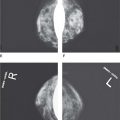
• Asymmetric breast tissue is usually identified when comparing one breast with the other. The breasts usually present a mirror image although 3%-5% of normal breast can show asymmetric densities in the outer quadrant or axillary tail.
• Areas of architectural distortion can represent a malignancy or a benign process such as surgical scars, sclerosing lesions, or posttraumatic fat necrosis.
• In-drawing or tenting of the breast parenchyma can be a sign of malignancy.
Calcification Morphology
• Calcifications within the breast may or may not be associated with a tumor, but should be evaluated separately from any associated tumor. Magnification mammography technique is extremely useful in evaluating calcification. Morphology as it relates to calcifications describes the form and structure of the calcifications, e.g., coarse, linear, round, casting, amorphous, or pleomorphic.
• Density—Low- or high-density radiopaque and any combination in between.
• Distribution—Placement of the calcifications within the breast.
• Change over time—Variation in density, distribution, number, morphology, or size over time.
• Number—Single or in clusters where a cluster of microcalcifications are described as 3-5 calcifications within an area no larger than 0.5-1 cm.
• Size—Varying from microcalcifications in the millimeter range to macrocalcifications in the centimeter range. Malignant-type calcifications are often microcalcifications.
• Coarse—Calcifications larger than 0.5 mm in diameter.
• Linear or rod-like—Calcifications over 1 mm in diameter and often associated with ducts.
• Round or punctuate—if the calcifications are smaller than 0.5 mm in diameter.
• Casting type—Fine, linear, or branching calcifications that can be fragmented with irregular contours and are often malignant.
• Amorphous or indistinct—Calcifications forming multiple flake-like irregular clusters that can be micro or macro and typically of intermediate concern.
• Pleomorphic or granular—Calcification of different shapes, irregular in form, size, and density and are typically malignant.
Benign Breast Calcifications
Appearance
• Smooth contours, high uniform density, e.g., plasma cell mastitis.
• Evenly scattered, homogenous, e.g., calcified arteries.
• Sharply outlined, spherical or oval, e.g., oil cysts.
• Pear-like densities resemble teacups or pearl drops on the lateral projection, e.g., milk of calcium.
• Bilateral and evenly scattered following the course of the ducts throughout much of the parenchyma, e.g., plasma cell mastitis.
• Ring-like, hollow, e.g., sebaceous gland calcifications.
• Eggshell-like, e.g., oil cyst, papilloma.
• Large bizarre size, e.g., hemangiomas.
• Milk of calcium is often due to cystic changes and can be associated with a micro cyst containing granules of calcified debris (milk of calcium). The calcifications are located within the lobules and because they are mobile, they will take the shape of the cavity where they are located. On the 90-degree lateral they will settle in the dependent portion of the lobules and are viewed as a crescent shape, resembling a teacup.
• Dermal calcifications are calcifications in the skin.
• Sutural calcifications are often seen in the breast after surgery and radiation therapy. Effects from radiation therapy may cause delays in the reabsorption of sutures. Calcium deposits on the sutures over time. These calcifications have a tubular appearance and are rarely seen if the patient does not have a history of radiation therapy.
• Vascular calcifications are calcified arteries or veins. Vascular calcifications are very distinctive and are seen mammographically as 2 parallel lines or broken tubular patterns.
• Oil cysts show mammographically as high-density tumors with lucent centers and eggshell-like calcifications. They usually form as a result of fat necrosis or are calcifying hematomas.
• Sclerosing adenosis or ductal hyperplasia is usually a result of increased cellular activity in the ducts and surrounding tissues. The condition can produce calcifications that tend to be linear and segmental but are sometimes malignant appearing.
• Ductal ectasia or plasma cell mastitis produces large calcifications that are the result of secretions within dilated ducts, whether periductal or intraductal. Periductal calcifications will have radiolucent centers representing the noncalcified centers of the duct. Because the calcifications are within the ducts they are linear and fragmental, forming along the long axis and pointing toward the nipple with only occasionally branching. Often these calcifications are bilateral and symmetrical in distribution.
Skin Thickening Syndrome
• Skin thickening can be the result of benign or malignant causes. Some causes include:
• Late malignancy either from direct invasion by the tumor or obstruction of the lymphatic or venous return.
• Inflammatory carcinoma will often result in diffuse unilateral skin thickening because there is direct tumor invasion of the dermal lymphatics.
• Infection and axillary lymphatic obstruction. These conditions can be secondary to breast carcinoma, metastases, or lymphomas.
• Radiation to the axilla.
• Cardiac failure or vena cava obstruction.
• Chronic renal failure.
Peau d’orange is used to describe generalized skin thickening. The skin will appear obviously thickened, most often in the lower dependent portion of the breast and takes on the appearance of an orange skin with prominent pores, hence the term—Peau d’orange. Mammographically, because the overall density of the breast is increased due to the high fluid content, there is a coarse reticular pattern on the mammogram.
MALIGNANT DISEASES
Location of the Breast Cancers
• The majority of breast diseases occur in the TDLUs; however, cancer can also grow in fibrous tissue, connective tissue, and larger ducts.
Common Breast Cancers
• The 2 main classifications of breast cancer are ductal and lobular carcinoma. Ductal carcinoma is the most common, accounting for about 90% of all breast cancers. Lobular carcinoma accounts for 5%-10% of all breast cancers.
• Ductal carcinoma in situ: The cancer is confined to the duct and does not invade the duct walls. There is a lot of controversy on the cancerous nature of this condition. This is commonly referred to as stage 0 carcinoma.
• Invasive or infiltrating ductal carcinoma: The cancer has spread from the ducts into the surrounding stromal tissue and may or may not extend into the pectoral fascia and muscle.
• Lobular carcinoma in situ is often not seen mammographically. The abnormal cells grow within the lobules but do not penetrate through the lobule walls. Lobular carcinoma in situ is sometimes referred to as lobular neoplasia and it is often considered to be a premalignant lesion that identifies women at an increased risk for subsequent development of invasive breast cancer.
• Invasive lobular carcinoma is often difficult to perceive on a mammogram and is better visualized using other modalities.
Other Breast Cancers
• Inflammatory, infiltrating medullary, mucinous or colloid, comedo, tubular, mucinous, papillary, and other breast cancers account for less than 10% of the total breast cancer cases and, apart from inflammatory carcinoma, they all have a better prognosis than infiltrating ductal or infiltrating lobular cancers.
Mammography Terminology
• Adenosis is enlargement and/or development of new lobular units.
• Apocrine metaplasia is a change occurring in the epithelial cells which exhibit characteristics of apocrine sweat glands.
• Biopsy is the pathologic examination of body tissue that has been removed from the body.
• Carcinoma is a malignant neoplasm or growth that can be visualized on a mammogram.
• Cysts occur in the TDLU when the extralobular terminal duct becomes blocked. The normal ductal secretions are not reabsorbed quickly enough and fluid begins to accumulate. Accumulating fluid causes pressure and the TDLU loses its shape as the cyst is formed.
• Duct ectasia is a benign process consisting of widened ducts containing thickened material. The cause is unknown.
• Epithelial hyperplasia describes an increase in the number of epithelial cells lining the duct. Certain stages can be considered premalignant.
• Fibroadenoma is a radiographically dense, encapsulated, round, and movable benign tumor seen frequently in women younger than 30 years. It develops from epithelial and fibroblastic tissue brought on by higher than normal estrogen levels.
• Fibrosis is the formation of fibrous tissue stemming from the connective and supportive stroma. Upon autopsy, up to 50% of all women have been found to have fibrocystic breasts.
• Fibrocystic changes represent development of benign cystic spaces within the ducts due to monthly hormone fluctuations that cause cysts to form throughout the breast tissue. Fibrocystic changes can mask or hide other malignant tumors.
• Fine-needle aspiration is a biopsy technique using a fine needle to aspirate or remove cells from a localized breast lesion for pathologic evaluation.
• Gynecomastia is the most common disorder that results in excessive development of the male breast from physiologic, hormonal, or pharmacologic causes.
• Hormone replacement therapy (HRT) is the prescribing of hormones for postmenopausal women to maintain their hormone levels to decrease symptoms and complications that could arise from declining estrogen levels.
• Intraductal papilloma is a benign, lobulated neoplasm, composed of benign epithelial tissue and located in the walls of ducts of the breast. It has a “berry-like” shape.
• Involution is the process of lobular regression. As a woman ages, the lobules will reduce in number and size with a decrease in the number of acini per lobule. The acini are the milk producing elements in the lobules. There is also replacement of the intralobular stroma with the dense collagen of breast connective tissue and over time there is progressive fatty replacement of the glandular elements.
• Lactation describes milk secretion from the breast.
• Lumpectomy is the removal of a tumor with excision of surrounding tissue. Lymph nodes are sometimes removed during the procedure.
• Mammography Quality Standards (MQSA) is the legislation passed by the United States Congress on October 1994, to ensure quality mammograms for all patients and to improve and standardize breast imaging.
• Mastectomy is the removal of the breast.
• Mastitis is inflammation of breast tissue.
• Mastorrhagia is the copious discharge of blood from the breast.
• Microcalcifications are minute calcium deposits in the breast that could possible indicate the presence of malignancy, although they can be benign.
• Multiparity is the condition of having carried more than 1 pregnancy past the point of viability (24-26 weeks of gestation) regardless of the outcome.
• Nulliparity describes a woman who has never given birth to a viable offspring. The nulliparous breast involutes more slowly than that of multiparous women and multiparity is generally associated with breasts that involute earlier.
• Parity is a condition of being pregnant and carrying a pregnancy past the point of viability or delivering a child.
• Postsurgical scarring can mimic a radial scar. It follows a surgical intervention and can appear as an area of architectural distortion or an irregular shaped lesion with spiculated margins. It may or may not be associated with calcifications. Typically, the surgical scar will resolve over time.
• Radial scar has a central fibrous core with radiating arms made up of benign epithelial growth and sclerosis. They are not truly scars and are often unrelated to prior surgery or trauma. Some possible causes of the radial scar are localized inflammatory reaction or chronic ischemia with a slow infection. The radial scar can be a benign condition, but can be associated with premalignant—atypical ductal hyperplasia—and malignant conditions.
• Sclerosing adenosis is adenosis with sclerosing of the intralobular stroma. This can present as calcifications or masses and is considered benign, and not a premalignant condition.
• Symmetry of the breast refers to breasts that are similar in size, shape, and composition of anatomy, so that one breast is a “mirror image” of the other.
Related posts:
Stay updated, free articles. Join our Telegram channel

Full access? Get Clinical Tree


