CHAPTER 5 ![]()
Diagnostic, Interventional, and Treatment Procedures
Summary of Important Points
INTERVENTIONAL OPTIONS
The first step in the fight against breast cancer is detection. However, after a lesion is discovered the next step is a histologic or cytologic analysis to definitely confirm or rule out malignancy. A finding can be palpable or nonpalpable, microcalcifications, mass densities, or an area of enhancement, but only by removing a sample of the tissue or cells in question and examining the removed sample using microscopic analysis, can a definitive diagnosis be made.
Cytologic Analysis
The microscopic examination of cell samples
Histological Analysis
The microscopic examination of tissue samples
Aspiration or FNA
Fine-needle aspiration (FNA) is removal of the content of a cyst for testing or analysis.
Stereotactic Breast Biopsy
• If a lesion is nonpalpable stereotactic imaging is an option to determine its exact location before it can be biopsied.
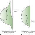
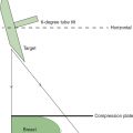
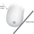
• The stereotactic equipment uses angled images to triangulate the depth of a lesion within the breast, and to calculate its position in 3 dimensions.
• The tube is angled 15 degree to the left and right along the × axis for 2 scout images, used for calculations of the depth (Z axis). The stereotactic calibrations are then used to position a biopsy probe within the breast at the calculated coordinates.
• This procedure can be performed either using a standard mammographic unit with an “add-on” attachment, or with a dedicated prone biopsy system. Some mammography units are capable of serving dual roles as an imaging unit and a biopsy system.
Advantages/Disadvantages of the Add-On Stereo Units
Advantages
• The patient is imaged in the upright position.
• The unit is relatively inexpensive when compared to the prone units.
• The units do not require a dedicated biopsy room.
• After use the unit can revert to use as a mammography unit.
• Imaging the posterior and axilla area of the breast is often easier using the add-on units.
Disadvantages
• There is less work space.
• The patient is upright with a greater risk of vasovagal reactions.
• Patient motion can be a problem.
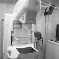
Advantages/Disadvantages of the Dedicated Prone Stereo Units
The prone units are dedicated procedure units where the patient lies prone with the breast of interest extending through a hole near the middle of the table. The mammography unit and needle guidance device are under the table. The table can be raised or lowered to suit the radiologist’s preference.
Advantages
• There is less chance for a vasovagal reaction.
• The patient lies in relative comfort during the procedure.
Disadvantages
• The unit is more expensive.
• Although these units can be used for needle localization, their major function as a biopsy unit will be underutilized if there is a low biopsy volume.
Preoperative Needle Localization for Excisional Biopsy
• If stereotactic technology is not available the preoperative needle localization technique can be used to locate any nonpalpable lesion or calcification within the breast. This procedure is done prior to a surgical excisional biopsy.
• The localization wire can be placed in the breast using either mammographic or sonographic guidance.
• Since this is a prelude to a surgical biopsy the patient should be scheduled for needle localization only after review of images and coordination of plans with a surgeon.
• The lesion is located and needle is placed directly in the lesion. It needle is removed leaving at least 5 mm of the wire guide within the lesion. The hook of the wire should be approximately 1 cm beyond the lesion.
• Any approach to the lesion should be parallel to the chest wall to minimize complications.
• In general, the shortest approach is used however sometimes an inferior approach will produce better cosmetic results.
• After the procedure, the relevant images, with the lesion clearly marked, should be sent to the surgeon. The wire should be carefully anchored in place after the localization because inaccurate placement or displacement of the wire can result in a missdiagonosis
• Some radiologists also provide the surgeon with line diagrams or a brief description, showing or telling the approximate distance from the breast skin surface to the lesion.
Breast Biopsy
Breast biopsy is the taking of a sample specimen for cytologic or histological analysis.
• The type of biopsy performed depends on a number of factors such as how suspicious is the abnormality, size, shape, and the location of the abnormality or the number of abnormalities present.
• Other relevant factors include the patient’s medical history, patient’s preference, training of the radiologist or surgeon, and the facility or equipment available.
Fine-Needle Biopsy
This technique is used to obtain cellular material from the area in question for cytologic analysis.
• Fine-needle biopsy (FNB) can be used to diagnose both cystic and solid lesions such as fibroadenomas. If the lesion is not palpable the needle can be guided to the lesion under ultrasound guidance or mammographically under stereotactic breast localization.
• FNB can reduce the necessity for a surgical breast biopsy but the accuracy of FNB is dependent on the individual performing the procedure—the radiologist or surgeon—and the ability of the cytologist.
Core Biopsy
• The core biopsy is the most commonly performed minimally invasive technique. The procedure can be performed under ultrasound, mammography, or MRI guidance.
• It is inexpensive, easy to perform, and highly accurate for many lesions.
• Core biopsy removes a sample of tissue versus FNB which removes only fluid and cells.
• The tissue samples are obtained using an 11-14 gauge or larger needle.
• Modality-based core biopsy is needed if a lesion is found only with 1 modality.
Automatic or Mechanical Core “Gun”
For this procedure the skin is numbed and a small cut (less than ¼ inch) is made. The gun needles have an inner needle with a trough extending within it. One end is covered by a sheath and attached to a spring-loaded mechanism. The device is inserted into the lesion. When the mechanism is activated, the needle moves forward, filling the trough with breast tissue. The outer sheath instantly moves forward to cut into the lesion and keep it in the trough. The gun-needle combination is designed to move a cutting needle rapidly through the breast. To yield adequate specimens a minimum of approximately 5 core samples are needed when assessing a lesion and at least 10 for an assessment of calcifications.
Vacuum Core Biopsy
For this procedure the skin is numbed and a small cut (less than ¼ inch) is made. A hollow probe is inserted and guided to the area of interest. A cylinder of tissue is then suctioned out and pulled into the probe through a hole in its side. A rotating knife inside the probe cuts the tissue sample from the rest of the breast.
Ultrasound Core Biopsy
Ultrasound can provide a highly accurate way to evaluate suspicious masses within the breast that are visible on ultrasound, whether or not they can be felt on the breast self-examination or the clinical breast examination. The procedure precludes the need to remove tissue surgically, plus it is faster than mammographically guided stereotactic biopsies and uses no ionizing radiation.
MRI-Guided Needle Biopsy
If a suspicious area is detected only on MR imaging or is not well seen on ultrasound or mammography, the radiologist may recommend an MR-guided breast biopsy. MR-guided biopsy requires only a topical anesthetic. Like ultrasound or mammographic minimally invasive biopsies, the MR biopsy is less costly than surgical biopsy, will leave little to no scarring, and can be performed in less than an hour.
Advantages of a Minimally Invasive Biopsy
• Can be performed as an outpatient procedure.
• Less visible scarring.
• Fewer complications and no anesthesia is involved.
• Less pain and usually less bleeding.
• Less costly since patients are sent home the same day and normally will not require an overnight stay in the hospital.
Open Surgical Biopsy
This is a major surgical procedure that will involve anesthesia and sometimes a hospital stay.
• Incisional biopsy is used to remove parts of a lesion.
• Excisional biopsy is used to remove the entire lesion.
Comparison of the Various Biopsy Methods
Complications and severity of side effects of a biopsy will depend on the method used and the quality of the care team.
• The open surgical biopsy has the lowest false-negative rates, less than 0.5% but is the most invasive and has the highest complication rate.
• The core biopsy has a reported 4%-10% false-negative rate but the most common error in core is understating the multifocality of the cancer, for example, a ductal carcinoma in situ could be diagnosed on a core but invasion found at excisional biopsy.
• FNB is less expensive, less invasive, and faster than core biopsy although it is more likely to lead to error due to insufficient sampling. The false-negative rate for the FNB can range from 5% to 20% and is higher for less experienced operators.
• The most substantial limitation for FNB is the high rate of insufficient sampling.
• Ultrasound-guided biopsy performed using any of the biopsy methods is faster and less expensive than stereotactic biopsy. It also does not involve ionizing radiation.
• Ultrasound also offers real-time visualization of the needle, which means the motion of the biopsy needle can be followed.
• Most palpable lesions are best biopsied under ultrasound guidance, whereas calcifications are best biopsied stereotactically with mammography guidance.
Specimen Radiography
The specimen is the breast tissue removed during a biopsy. A specimen radiograph should be performed after every biopsy to confirm that the lesion was removed and that the margins are clean.
• In imaging the specimen, speed, and efficiency are important since the patient may be under anesthesia.
• The mammographer should always use compression and magnification is recommended for calcifications.
• If microcalcifications are present in a specimen they should be counted and noted.
• The radiologist should indicate where the pathology is located on the specimen, as this will help the pathologist in evaluating the lesion.
• If the tumor is close to the margins of the specimen or if the margins are positive for cancer, additional tissue must be excised before the incision is closed.
Ductography or Galactography
• Ductography can be performed to evaluate a suspicious nipple discharge. The discharge can be spontaneous, bilateral or unilateral and may be clear, serous, or bloody.
• The technique can only be performed if nipple discharge is present.
• Ductography can detect abnormalities such as filling defects, wall irregularity, duct expansion, or abrupt cutoff of a duct; however, the procedure is rarely performed today because ultrasound can now be used to identify and localize duct abnormalities or filling defects.
Pathology Review
• The final stage of any interventional process is the pathology review. There should be good communication among the radiologist, the surgeon, and the pathologist.
• Most facilities now have a system in place to track all positive pathologic findings.
• A tracking system is required to comply with the Mammography Quality Standards Act (MQSA).
• Regular reviews and medical audits will help the facility to identify areas of concern including high false-negative or false-positive rates. By identifying possible problem areas, the facility can improve service and enhance breast cancer detection and screening.
Mammoplasty
• Mammoplasty is the general term used when describing reshaping of the breast. The breast can be lifted to reduce a sagging breast, enlarged (augmented), or reconstructed after the removal of a tumor.
Cosmetic Interventional
• Cosmetic interventional includes any reconstruction of the breast that is not related to medical reasons. This can include reduction or augmentation. Over the years, breasts will lose their shape and firmness from pregnancies, nursing, and the aging process. Cosmetic intervention can improve self-confidence or improve appearance.
• Surgery and reconstruction after breast cancer does not fall under the heading of cosmetic intervention.
Breast Reduction
• The process reduces the overall size of the breast. It can be done for cosmetic or medical reasons. Some of the breast tissue is removed and the nipple area often relocated higher on the breasts for better cosmetic results.
Breast Augmentation
• A technique to increase breast size. It can be performed for cosmetic or medical reasons.
• Implants can be placed in front of the pectoral muscle (subglandular or retromammary implants) or behind the pectoral muscle (subpectoral or retropectoral implants).
BREAST CANCER TREATMENT OPTIONS
Surgical Options
Mastectomy is the surgical removal of the entire breast. There are different types of mastectomy.
• Radical mastectomy removes the entire breast, lymph nodes, and chest wall muscles under the breast. It is rarely performed today because the modified mastectomy is just as effective plus the modified is less debilitating and deforming.
• Modified radical mastectomy removes the entire breast including the nipple/areolar region and some of the underarm lymph nodes.
• Prophylactic mastectomy removes a healthy breast when the individual has a high risk factor for developing breast cancer. All the breast tissue including the nipple/areolar region may be removed. Alternatively, nipple sparing mastectomy may be performed.
• Lumpectomy removes the breast cancer tumor and surrounding margins of the normal breast tissue.
• Lumpectomy is often combined with other therapy options such as radiation treatment, chemotherapy or hormonal treatment.
• If followed by radiation therapy, the patient is usually given 6 weeks of radiation therapy to ensure that all cancer cells in the remaining breast have been destroyed. Radiation treatment usually begins 1 month after surgery after giving the breast time to heal. However, there are newer radiation treatment options that begin and end earlier.
• Lumpectomy can be followed by chemotherapy to control the systemic spread of breast cancer or treatment with the drug Tamoxifen.
• Breast conservation should only be performed if the treatment provides a cure rate equal to that obtained from a mastectomy.
Surgical Complications
• Wound infection.
• Hematoma or microhematoma.
• Seroma—clear fluid trapped in the wound.
• Lymphedema if the lymph nodes are removed during the operation.
• Numbness in the upper arm and skin—can be caused by the removal of lymph nodes and the resulting edema.
• Phantom breast pain/sensations. These symptoms can include unpleasant itching, pins and needles, pressure and throbbing often associated with small nerves between the breast tissue and skin, which were cut during surgery. The neural connection in the brain is undergoing a process of reorganization called neural plasticity. This results in the spontaneous firing of electrical signals from the ends of the cut or injured nerves causing the phantom sensations. Phantom pain can be relieved by massage and or pain medication.
Axillary Node Dissection
This procedure removes the underarm lymph nodes during a mastectomy or lumpectomy. A common complication of axillary node dissection is lymphedema or chronic swelling of the arm due to the accumulation of lymphatic fluids. Lymphedema is exaggerated when axillary node dissection is combined with radiation therapy.
Sentinel Lymph Node Biopsy
This is a more conservative process and involves the removal of only 1-3 sentinel lymph nodes in the axillary area. Prior to a sentinel node biopsy the sentinel node or first nodes in the lymphatic chain must be identified using any of the adjunctive detection options. If cancer is discovered in the sentinel nodes more nodes are usually removed.
Breast Cancer Staging
Staging determines the nature of the cancer whether it is invasive or noninvasive including its size, its location, if there is lymph node involvement and if the cancer has spread to other parts of the body. Staging results are important in determining prognosis and treatment options. The American Joint Committee on Cancer (AJCC) has a standardized staging system based on “T,” “N,” and “M” where “T” is related to the tumor size, “N” denotes lymph node spread, and “M” assesses distant metastasis.
RADIATION THERAPY
Radiation treats breast cancer with high-energy radiation to destroy cancer cells in the chest wall, axilla, and the breast. Radiation therapy is often used in conjunction with other treatment options to remove the remaining cancer cells in the breast, chest wall, and axilla area and can also be used before surgery to shrink the size of the tumor.
External beam radiation usually begins 1 month after surgery after giving the breast time to heal. Treatment time is typically 15-30 minutes given 5 times per week for 5-8 weeks. The procedure itself is painfree and the patients are generally outpatients. During treatment patients will be monitored via closed circuit television.
Internal Radiation
Internal beam radiation or brachytherapy involves applying the radiation from the inside out. Treatment can be reduced from 8 weeks to 5-9 days.
Advantages of Internal Beam Radiation (Brachytherapy)
• Reduction in the time of treatment from 6 weeks to 7-9 days.
• Less irritation of healthy breast tissue.
• Less delay before the start of treatment—the treatment can start immediately after a lumpectomy.
• Fewer skin reactions such as redness, rashes, or irritation.
Side Effects of Radiation Treatment
Most side effects will disappear after 6-12 months. The breast will sometime remain firm. On rare occasions the breast may become enlarged due to fluid buildup or smaller due to tissue changes. The skin can become more or less sensitive. Other side effects can include:
• Fatigue—extreme tiredness.
• Swelling of the breast.
• Heaviness in the breast.
• Sunburn-type appearance of the breast skin.
• Loss of appetite.
DRUG AND HORMONAL OPTIONS
Chemotherapy is an adjuvant therapy and involves the use of drugs to treat cancer that may have spread beyond the breast. Neoadjuvant chemotherapy is given before surgery to help shrink the cancerous tumor. Adjuvant chemotherapy is given in addition to another breast cancer treatment, for example, mastectomy.
• Chemotherapy regimens are tailored for the individual patient and can vary tremendously. The type of treatment will depend on the patient’s age, overall health, stage and grade of the cancer, past or future treatment, and other health problems.
• Chemotherapy is often a combination of drugs. Most physicians prefer a combination of lower doses of individual drugs versus a high dose of 1 powerful drug. Lower dose drugs are also associated with fewer side effects.
• Chemotherapy is a systemic treatment and because the drug is distributed throughout the body it will affect all the cells, tissues and organs in the body. Many chemotherapy drugs work to kill actively reproducing cells; however, some drugs will work on cells in a particular phase of the cell cycle.
Method of Chemotherapy Administration
• The chemotherapy is often administered through a semipermanent catheter (port-a-cath) implanted into a large vein in the arm or hand or the subclavian vein near the clavicle. Some chemotherapy drugs are given orally as tablets or liquids. Chemotherapy can also be given as an intramuscular injection, topically, or injected directly into the cancer area.
• Chemotherapy courses can be given daily, weekly, monthly, or other scheduling options depending on the patient’s response to the drug.
• The treatment lasts 3-6 months. Most long chemotherapy sessions include built-in rest cycles to give the healthy cells recovery time.
Hormonal Treatment
This often involves the use of a class of drugs called selective estrogen receptor modulators or SERMs. They are used to shrink or stop the recurrence of breast cancer or to lower the risks of breast cancer recurrence in postmenopausal women.
Tamoxifen Treatment
• Tamoxifen is an antiestrogen drug that blocks estrogen and can lower the risk of breast cancer recurrence in postmenopausal women after surgery.
• Some cancers have estrogen receptors (ER positive) on the surface of their cells. These cancers rely on a supply of estrogen to grow. Tamoxifen prevents estrogen from latching onto tumor cell receptors and directing them to multiply.
• Tamoxifen is also used to shrink large tumors.
• Tamoxifen by itself is recommended for 5 years. Some studies now suggest that it offers added protection after 5 years. The benefits of Tamoxifen are thought to outweigh the risks.
Rolaxifene
• Works similar to Tamoxifen but is less toxic.
Herceptin
• Can be used to treat HER2-positive metastatic breast cancer.
Aromasin
• Carries fewer side effects than Tamoxifen. Aromasin suppresses blood levels of estrogen by blocking aromatase, the enzyme that helps the body produce it.
Pain medication is available for patients in severe pain from cancer. Newer pain medications are even more potent than morphine.
Postbreast Cancer—Breast Reconstruction
Breast reconstruction aims to restore the appearance of the breast.
• Options include reconstruction using saline or silicone implants. Modern techniques (subpectoral or retropectoral placement) place the implants behind the pectoral muscle. Older techniques (subglandular or retromammary placement) placed the implants in front of the pectoral muscle.
• Flap surgery removes skin and fat from the abdomen, back, or buttock and uses it to form a new breast. Flap techniques can be pedicle flap, where the flap remains attached to its original blood supply and it is tunneled under the skin to the breast area, or free flap, where the flap is completely removed and microsurgery is needed to recreate a blood supply when the flap is positioned as a breast mold. The recovery time for most flap reconstruction is 4-6 week. Patients are usually required to have a drain for 1-2 weeks.
Questions
1. The surgical specimen is radiographed to
(A) confirm that the entire lesion was removed
(B) compare various needle localization techniques
(C) magnify the lesion to assess any possible microcalcifications
(D) check the position of the lesion
2. The specimen is compressed to reduce
(A) motion unsharpness
(B) radiation exposure
(C) tissue thickness
(D) magnification factor
3. Which procedure is performed to obtain cellular material from a suspicious area for cytological analysis?
(A) ductography
(B) needle localization
(C) pneumocystogram
(D) fine-needle biopsy (FNB)
4. Preoperative localization will
1. direct the surgeon to the area requiring biopsy
2. help the surgeon to excise a smaller specimen
3. ensure that the correct area is removed
(A) 1 and 2 only
(B) 2 and 3 only
(C) 1 and 3 only
(D) 1, 2, and 3
5. Core biopsy techniques developed as an alternative to surgical biopsy because this technique provided a larger sample of the area of suspicion and thus more information than
(A) ductography
(B) needle localization
(C) pneumocystogram
(D) FNB
6. An ultrasound of a lesion showed a spherical mass with smooth regular borders, anechoic interior, and acoustic enhancement. The lesion is likely to be a
(A) fibroadenoma
(B) abscess
(C) simple cyst
(D) ductal carcinoma
7. In ultrasound, the term acoustic enhancement refers to
(A) a structure without internal echoes
(B) a structure with internal echoes
(C) the amount of sound passing through an anechoic structure
(D) few echoes within a structure
8. A procedure whereby the lactiferous duct is cannulated and a small amount of contrast agent is injected into the duct is termed
(A) ductography
(B) needle localization
(C) pneumocystogram
(D) FNA
9. A patient had an ultrasound, which confirmed the presence of a cyst in the breast. The radiologist wished to assess the contents of the cyst. What additional study could be recommended?
(A) ductography
(B) needle localization
(C) pneumocystogram
(D) FNA
10. Biopsy performed using a 14-gauge needle to remove tissue samples from the breast is termed
(A) core biopsy
(B) cytology
(C) ductography
(D) aspiration
11. Stereotactic breast localization is used to
(A) obtain 2-dimensional information on palpable breast lesions
(B) calculate the vertical position of nonpalpable lesions
(C) obtain a 3-dimensional image of the breast
(D) calculate the horizontal, vertical, and depth position of nonpalpable lesions
12. A cyst aspiration can only be performed
(A) under ultrasound guidance
(B) using mammographic imaging
(C) using MRI guidance
(D) none of the above
13. Which of the following biopsy techniques is most accurate?
(A) FNB
(B) open surgical biopsy
(C) core biopsy
(D) MRI core biopsy
14. In general, the optimal duration of Tamoxifen treatment is
(A) 2 years
(B) 3 years
(C) 4 years
(D) 5 years
15. Lumpectomy describes the process of
(A) removing the entire breast including the nipple
(B) removal of the breast cancer tumor and surrounding margins of normal breast
(C) making a small incision over or near the site of the breast lesion
(D) removing benign lumps from the breast
16. Radiation therapy can be used
1. with other treatment options
2. to kill any remaining cancer cells in the breast, or chest wall area
3. to shrink the size of a tumor before surgery
(A) 1 and 2 only
(B) 2 and 3 only
(C) 1 and 3 only
(D) 1, 2, and 3
17. Chemotherapy is a class of drugs that can be used to
(A) kill cancer cells in other parts of the body
(B) block estrogen from cancer cells
(C) reduce estrogen levels in the body
(D) kill cancer cells by using high-energy radiation
18. Breast reconstruction can involve the placement of small fluid-filled sacs behind the pectoral muscle. Two common types of such implants are
(A) saline and flap surgery
(B) TRAM flap and silicone implant
(C) silicone or saline implants
(D) saline-filled implant and latissimus dorsi flap implant
19. Chemotherapy
(A) involves the use of drugs to treat cancer that may have spread
(B) is the destruction of cancer cells using estrogen receptor drugs
(C) involves mapping the area around a tumor with the injection of a radioactive tracer
(D) is the removal of the cancerous tissue from the breast
20. A process of removing tissue and fat from the abdomen and transferring that tissue to reconstruct the breast is called
(A) deep inferior epigastric perforators (DIEP) flap
(B) latissimus dorsi flap
(C) TRAM flap
(D) TUG flap
21. Antiestrogen drugs such as Tamoxifen can be used to
1. slow or stop the cancer’s growth
2. prevent breast cancer in high-risk women
3. prevent the recurrence of breast cancer
(A) 1 and 2 only
(B) 2 and 3 only
(C) 1 and 3 only
(D) 1, 2, and 3
22. MRI imaging
(A) uses complex magnetic properties of elements
(B) explores the behavior of liquids or solids in metal
(C) images the patient in less than 15 minutes
(D) is less sensitive than a mammogram
23. Most of the risk of MR imaging of the breast are associated with
(A) the magnetic properties of the patient
(B) ferromagnetic metals
(C) technologist errors
(D) radiologist errors
24. In breast cancer staging the patient was rated N1. This means that
(A) the cancer has not spread beyond the breast
(B) the cancer is confined to 1-2 lymph nodes
(C) the patient has a small tumor
(D) the is no evidence of metastasis
25. Cyst aspiration describes the removal of
(A) a sample of the content of a cyst for biopsy
(B) cell samples taken from a cyst for analysis
(C) fluid from a cyst
(D) any mass from the breast
26. If a breast lesion is seen only on the breast ultrasound, which modality should be used when performing a biopsy of the lesion?
(A) mammogram
(B) ultrasound
(C) MRI
(D) nuclear medicine
27. The pathology review is
(A) a method of tracking and reviewing all positive findings
(B) a process of eliminating all positive findings
(C) a method of tracking cosmetic intervention
(D) a process of identifying all mammoplasty
28. The technique used where by contrast is injected into the ducts to check for an abnormality is called
(A) augmentation
(B) ductography
(C) biopsy
(D) mammoplasty
29. Cosmetic intervention could be considered
(A) breast reconstruction after treatment for breast cancer
(B) surgical reconstruction of the breast to increase its firmness
(C) breast biopsy to check a suspicious lesion
(D) breast reduction to relieve back pain
30. A process by which the breast size is increased using implants is called
(A) augmentation mammoplasty
(B) ductography
(C) needle biopsy
(D) needle aspiration
31. The removal of 1-3 axillary nodes for testing is included in a/an
(A) axillary node dissection
(B) sentinel node mapping
(C) sentinel node biopsy
(D) lumpectomy
32. Specimen radiography confirms that
(A) a biopsy is not necessary
(B) the patient has no malignancy
(C) the patient needs a mastectomy
(D) the suspected lesion is in the tissue that was removed
33. Which of the following are side effects of radiation treatment?
1. loss of appetite
2. fatigue
3. heaviness of the breast
(A) 1 and 2 only
(B) 2 and 3 only
(C) 1 and 3 only
(D) 1, 2, and 3
34. A big advantage of brachytherapy over traditional radiation therapy treatments is
(A) reduced time of treatment
(B) the ability of brachytherapy to treat larger tumors
(C) more radiation can be given to healthy tissue
(D) it is more convenient because of the longer treatment times
35. Side effects of chemotherapy include
(A) nausea and or vomiting
(B) reduced white cell count
(C) none of the above
(D) A and B
36. What is Tamoxifen?
(A) an adjuvant therapy using drugs to reduce the risk of certain cancers
(B) a drug regimen that will effectively block hormones in the body
(C) an external method of killing cancer cells
(D) the removal of all cancer cells from the body thereby reducing cancer risks
37. Which of the following biopsy techniques is the most accurate?
(A) FNB
(B) core biopsy
(C) stereotactic breast biopsy
(D) open surgical biopsy
38. Which of the following is the least expensive and least invasive method of breast biopsy?
(A) FNB
(B) core biopsy
(C) open surgical biopsy
(D) stereotactic breast biopsy
39. A technique used to assess and evaluate nipple discharge by checking for defects or wall irregularity is called
(A) ductography
(B) interventional radiography
(C) aspiration biopsy
(D) lumpectomy
40. Neoadjuvant chemotherapy refers to treatment
(A) before surgery to help shrink the size of cancerous tumors
(B) after mastectomy to slow the growth of cancer
(C) before surgery utilizing an antiestrogen drug
(D) after surgery that involves radiation
41. Preoperative localization is
(A) stereotactic breast localization of a palpable abnormality
(B) used to locate palpable breast lesions
(C) a technique use to locate nonpalpable lesions
(D) a check of the tissue for malignancy
42. A biopsy technique that often requires the presence of a cytologic technologist describes
(A) open surgical biopsy
(B) core biopsy
(C) stereotactic breast biopsy
(D) fine-needle biopsy
43. Cosmetic intervention describes
1. breast reconstruction after treatment for breast cancer
2. surgical reconstruction of the breast at the patient’s request
3. breast augmentation for personal reasons
(A) 1 and 2 only
(B) 2 and 3 only
(C) 1 and 3 only
(D) 1, 2, and 3
44. The side effects of chemotherapy are related to the effect of the drug on
(A) all tissues and organs in the body
(B) cancer cells only
(C) the growth of cancer
(D) hair follicles only
45. Any size tumor with cancer spread to chest wall and/or skin of breast and up to 10 axillary or claviclar lymph nodes is describes as stage
(A) 1
(B) 2
(C) 3
(D) 4
46. Which of the following would be contraindicated for lumpectomy?
1. women with previous lumpectomy
2. women with multiple cancer sites in 1 breast
3. large tumors in small breast
(A) 1 and 2 only
(B) 2 and 3 only
(C) 1 and 3 only
(D) 1, 2, and 3
47. The staging of a cancer will determine
1. if the cancer is invasive
2. the exact tumor size
3. if the cancer has spread to distant sites
(A) 1 and 2 only
(B) 2 and 3 only
(C) 1 and 3 only
(D) 1, 2, and 3
48. In breast cancer staging the patient was rated T1. This means that
(A) the cancer has not spread beyond the breast
(B) the cancer is confined to 1-2 lymph nodes
(C) the patient has a small tumor
(D) there is no evidence of metastasis
Related posts:
Stay updated, free articles. Join our Telegram channel

Full access? Get Clinical Tree


