Aneurysmal Bone Cyst


Clinical Presentation


The patient is a 10-year-old female with a 7-month history of upper back pain, scapular pain, and rib pain. The patient developed bilateral lower extremity weakness 3 weeks prior to presentation.


Imaging Presentation


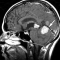
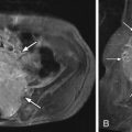
Magnetic resonance (MR) imaging reveals a solid and cystic mass containing heterogeneous regions of high, low, and intermediate signal intensity ( Fig. 7-1 ) . The mass predominantly involves the posterior vertebral arch of T3 on the left, with some involvement of the T3 vertebral body on the left. Computed tomography (CT) shows a large soft tissue mass that has destroyed the T3 spinous process, left lamina, and left pedicle, and extends into the vertebral body ( Fig. 7-2 ) .


Figure 7-1


T3 Vertebral Aneurysmal Bone Cyst.

T2-weighted left parasagittal MR image A and mid sagittal image B . The aneurysmal bone cyst predominantly involves the posterior vertebral arch of T3 ( short arrow ). The lesion contains multiple ovoid areas of heterogeneous signal intensity that has replaced the left posterior vertebral arch and left posterolateral aspect of the vertebral body ( long arrow ). At least one loculation ( thin arrow in image A ) contains a fluid/fluid level, which is vertical on the image with the patient supine. The lesion extends into the epidural space causing cord compression. The lesion was largely cystic in nature at surgery, containing multiple loculated cystic collections of bloody liquid.


Figure 7-2


T3 Vertebral Aneurysmal Bone Cyst.

Contrast enhance axial CT image. Minimal contrast enhancement is seen along the margin of the lesion ( arrows ). The lesion was almost entirely cystic in nature at surgery containing multiloculated bloody liquid at different stages of metabolic degradation. Ovoid hypodense areas with the lesion represent lower density liquid-filled cystic cavities. More dense areas within the lesion represent cystic cavities containing more proteinaceous bloody liquid.


Discussion


The lesion that has become known as “aneurysmal bone cyst” was originally described by Van Arsdale in 1893, who referred to this lesion as a “ossifying hamartoma.” The term aneurysmal bone cyst was first used for describing these lesions in 1942 by Jaffe and Lichtenstein. Aneurysmal bone cysts are rare, benign, yet locally destructive; they are expansile, highly vascular bone lesions. They represent approximately 1% to 2% of primary bone “tumors,” although they are not true neoplasms. The majority of aneurysmal bone cysts arise in the metaphysis of the long bones. Approximately 10% to 20% of aneurysmal bone cysts occur in the axial skeleton. Spinal aneurysmal bone cysts occur most frequently in the thoracic spine, followed by the lumbar spine, cervical spine, and least commonly in the sacrum. Aneurysmal bone cysts in the sacrum or pelvis tend to be more aggressive lesions that cause extensive bone destruction ( Figs. 7-2 to 7-8 ) . Spinal aneurysmal bone cysts nearly always arise in the posterior vertebral arch but frequently extend into the ipsilateral pedicle and vertebral body, epidural space, or adjacent neural foramen (see Figs. 7-1 and 7-2 ). They rarely extend into the nearby ribs or adjacent vertebrae.


Figure 7-3


Sacral Aneurysmal Bone Cyst.

25-year-old male patient with 2-month history of low-back pain and hamstring distribution pain. AP radiograph of sacrum shows a subtle Iow a density “bubbly lesion” throughout the sacrum ( arrows ), which is slightly less dense compared to the normal density of the iliac bones. This lesion could easily be overlooked on this radiograph, possible mistaken for overlying bowel.


Figure 7-4


Sacral Aneurysmal Bone Cyst.

Same patient as in Figure 7-3 . Axial CT Image reveals a homogeneous hypodense lesion ( arrows ) that occupies almost the entire sacral contents, sparing the cortical margins of the sacrum. The sacroiliac joints and adjacent cortex remains intact.


Figure 7-5


Sacral Aneurysmal Bone Cyst.

Axial T1 weighted MR image corresponding to same slice location as CT image in Figure 7-4 . The sacrum ( arrows ) is uniformly hypointense relative to the iliac bones.


Figure 7-6


Sacral Aneurysmal Bone Cyst.

Same patient as in Figure 7-5 . Corresponding T2 weighted MR axial image. The sacral contents are almost entirely filled by homogeneously T2 hyperintense liquid ( arrows ).


Figure 7-7


Sacral Aneurysmal Bone Cyst.

Same patient as Figures 7-4 to 7-6 . Contrast enhanced fat saturated coronal T1-weighted MR image. There is faint enhancement of the cystic contents within the sacrum. The margins of the cyst ( arrows ) enhance more intensely than the central portion of the cyst.


Figure 7-8


Sacral Aneurysmal Bone Cyst.

Same patient as in Figures 7-3 to 7-7 . Following sacral curettage, the sacral cavity was packed with morsellized bone allograft resulting in heterogeneous hyperdense appearance of the sacrum ( arrows ) on this axial postoperative CT scan. Compare with preoperative CT scan, Figure 7-4 .


The majority of patients who present with aneurysmal bone cyst are younger than age 20, and there is a slight female predilection; 95% of patients with spine lesions present with back pain, usually slow in onset. These patients may have scoliosis. Myelopathy or radiculopathy may be present if the spinal cord or nerve roots are compressed. Paresthesias, loss of anal sphincter tone, leg weakness, and urinary retention may occur with spinal or sacral aneurysmal bone cysts depending on their location and extent.


Secondary aneurysmal bone cysts may arise in or adjacent to other primary bone tumors in a small percentage of cases, especially in giant cell tumors and osteoblastomas, but they also occasionally occur in osteosarcomas, chondroblastomas, fibrous histiocytomas, and less commonly in other bone tumors. The etiology of aneurysmal bone cysts is unknown. It is possible that some of these tumors arise secondary to vascular anomalies such as arteriovenous (AV) fistula formation in bone secondary to trauma or neoplasm. Hereditary influences are believed to play a role in a significant number of primary aneurysmal bone cysts. Translocations or deletions in chromosomes 16 and 17 have been demonstrated in some patients.


Histologically, the typical aneurysmal bone cyst is composed of blood-filled cavities with surrounding trabecular and solid elements comprised of vascularized stromal tissues containing multinucleated osteoclastic giant cells, spindle cells, fibroblasts, myofibroblasts, hemosiderin deposits, and a variable amount of cartilage and osteoid that may be mineralized.

Only gold members can continue reading. Log In or Register to continue

Stay updated, free articles. Join our Telegram channel

Aug 25, 2019 | Posted by in NEUROLOGICAL IMAGING | Comments Off on Aneurysmal Bone Cyst

Full access? Get Clinical Tree

Get Clinical Tree app for offline access