Appendicitis
Kelly L. Hastings
Cassandra M. Sams
CLINICAL HISTORY
35-year-old male with right lower-quadrant abdominal pain.
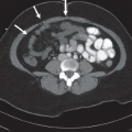
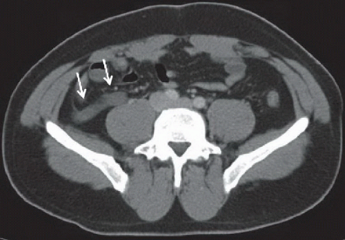 FIGURE 63A |
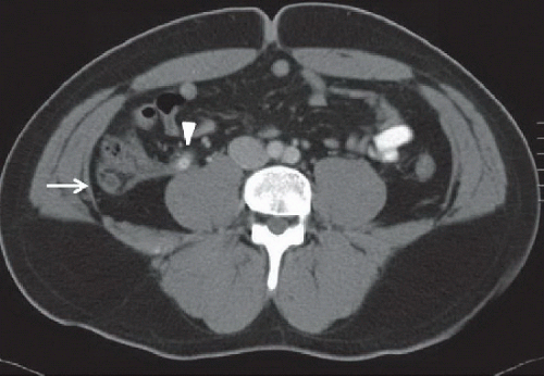 FIGURE 63B |
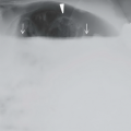
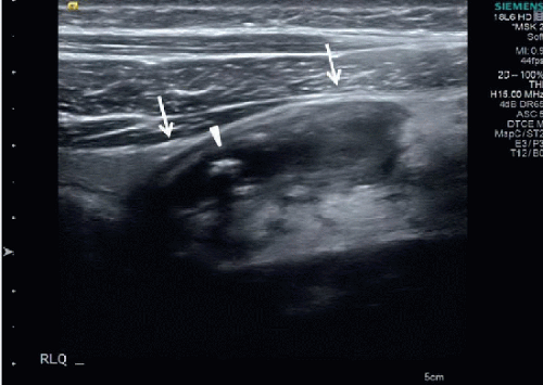 FIGURE 63C |
FINDINGS
Figure 63A: Axial contrast-enhanced CT scan of the appendix. The appendix is dilated and fluid-filled (arrows) with periappendiceal stranding. Figure 63B: Axial contrast-enhanced CT scan of the appendix of the same patient. The distal appendix is dilated and fluid-filled with appendiceal wall enhancement and periappendiceal stranding (arrow). An appendicolith is identified in the proximal appendix (arrowhead). Figure 63C: Grayscale ultrasound image of the right lower quadrant of a different patient shows a blind-ending, dilated tubular structure with thickened wall, consistent with a dilated, edematous appendix (arrows). Hypoechoic material fills the appendix. An echogenic round structure in the midappendix demonstrates posterior shadowing, compatible with an appendicolith (arrowhead). These findings are consistent with acute appendicitis.
Related posts:


Stay updated, free articles. Join our Telegram channel

Full access? Get Clinical Tree


