CHAPTER 72 Arterial Anatomy of the Abdomen
NORMAL ANATOMY
General Anatomic Descriptions
The abdominal aorta and its branches are responsible for the arterial blood supply to the abdominal organs (Figs. 72-1 and 72-2). From the thoracic cavity, the aorta courses through the diaphragm slightly to the left of midline at the aortic hiatus, most commonly anterior to the twelfth thoracic vertebral body.1 The normal aorta measures 1.5 to 2 cm in diameter and diminishes in caliber until its terminal bifurcation, typically at the level of the fourth lumbar vertebral body. During the process of vasculogenesis, embryonic folding during the fourth week parallels fusion of the paired dorsal aortae into a single median dorsal aorta in the abdomen.2 Major branches from the dorsal abdominal aorta subsequently develop ventrally, laterally, and posterolaterally and may be divided into four groups (Table 72-1).
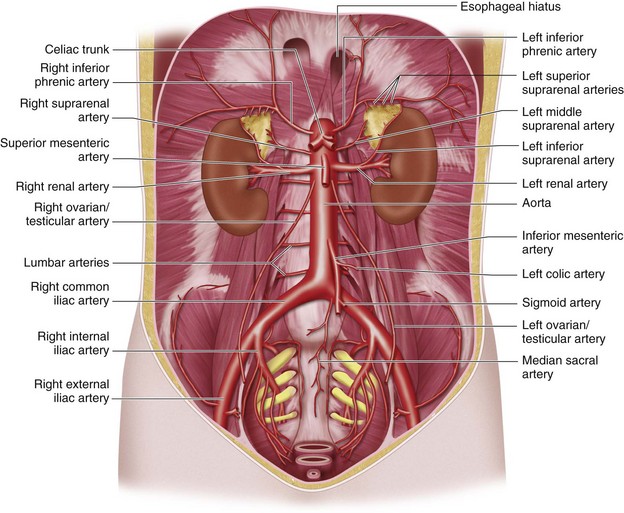
 FIGURE 72-1
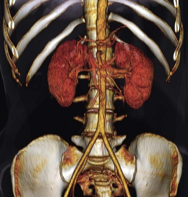
 FIGURE 72-2
TABLE 72-1 Major Branches of the Abdominal Aorta
| Group | Arterial Branches |
|---|---|
| Group 1 | |
| Group 2 | |
| Group 3 | |
| Testes and ovaries | Testicular and ovarian arteries |
| Group 4 | |
| Terminal and posterior branches | |
Group 1 includes branches formed by the union of vitelline arteries arising from the wall of the yolk sac that supply organs depending on their location in the primitive gut.2 The developing gastrointestinal tract, or primitive gut, is often divided into the foregut, midgut, and hindgut. The foregut gives rise to the esophagus, stomach, proximal half of the duodenum, liver, and pancreas. The midgut gives rise to the distal half of the duodenum, jejunum, ileum, cecum, appendix, ascending colon, and the right two thirds of the transverse colon. The hindgut gives rise to the left two thirds of the transverse colon, descending colon, sigmoid colon, and rectum. From the dorsal aorta, the three dominant vitelline arteries are further refined into the celiac trunk, superior mesenteric artery, and inferior mesenteric artery, which correspond to the three primitive gut regions, respectively.
Group 2 (renal and adrenal branches) includes branches supplying the kidneys and adrenal glands.
The arterial supply of the pelvic organs and pelvic wall courses through the internal iliac arteries, which originate at the bifurcation of the common iliac artery at the sacroiliac junction. Arterial anatomy of the pelvis is presented in Chapter 73.
Detailed Description of Specific Areas
Normal Variants
Group 1: Celiac Trunk and Mesenteric Arteries
The celiac trunk is a short (1 to 2 cm) ventral branch of the abdominal aorta arising below the aortic hiatus, typically between T12 and L1 (Fig. 72-3).1,3,4 The foregut organs are supplied by the celiac trunk’s trifurcated branches, the left gastric, common hepatic, and splenic arteries, in up to 75% of individuals (Fig. 72-4). Rarely, a common origin of the celiac and superior mesenteric artery develops; however, most variants involve separate origins of one or more of the three main celiac branches. The left gastric artery is ordinarily the first and smallest celiac branch, supplying the distal esophagus and stomach. Coursing along the lesser curvature of the stomach, the left gastric branches unite with the right gastric branches, forming a vascular arcade. Further anastomoses exist between the left gastric artery and the short gastric arteries from the splenic artery as well as the left gastroepiploic (sometimes called gastro-omental) artery.
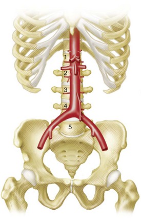
 FIGURE 72-3
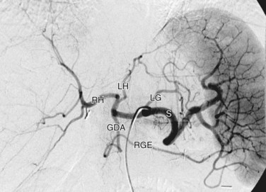
 FIGURE 72-4


