Fig. 32.1
Arterial anatomy of the upper extremity
1.2 Venous (Fig. 32.2)

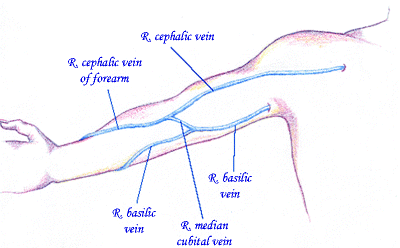
Fig. 32.2
Superficial venous anatomy of the upper extremity
Superficial
The superficial venous system consists of the cephalic vein, which runs laterally from the wrist to its insertion into the axillary vein, and the basilic vein, which runs posterior and medial throughout the forearm crossing anterior to the antecubital fossa to its insertion into the axillary vein.
Deep
The deep venous system usually is paired and courses parallel to the arteries; it consists of the superficial and deep palmar arch, which form to produce the radial and ulnar veins, which combine to produce the brachial followed by the axillary and subclavian vein. The main variation from arterial anatomy is proximal; both the right and left subclavian vein combine with their respective internal jugular veins, to form both a right and left innominate (brachiocephalic) vein; the right and left innominate vein combine to form the superior vena cava.
2 Disease Definition
The National Kidney Foundation-Dialysis Outcomes Initiative (NKF-KDOQI) recommends that patients be referred to a vascular access surgeon for permanent dialysis access when their creatinine clearance is less than 25 mL/min.
3 Disease Distribution
In 2005, data from the U.S. Renal Data System (USRDS) showed more than 106,000 new patients began therapy for end-stage renal disease (ESRD) while the prevalent dialysis population reached 341,000. Total Medicare costs for ESRD patients neared $20 billion; this accounts for 6% of the total Medicare spending in 2005.
4 Classification of Treatment Modalities
4.1 Dialysis Catheters
Short-term dialysis catheters are double lumen noncuffed nontunneled catheters that are placed at beside without fluoroscopic guidance; these catheters are placed in patients needing acute dialysis access and are used for less than 3 weeks duration. Long-term dialysis catheters are double lumen cuffed tunneled catheters that are placed with fluoroscopic guidance; these catheters are placed in patients needing long-term dialysis access usually while awaiting permanent arteriovenous (AV) access and are intended to be used over weeks to month’s duration. Both short- or long-term dialysis catheters may be placed in the internal jugular, subclavian, or femoral veins; for highest flow and lowest complications, the right internal jugular approach with the catheter tip just above the atrial-caval junction is preferred. To decrease the risk of central venous stenosis and subsequent venous hypertension, the subclavian approach is avoided if at all possible.
4.2 Autogenous Arteriovenous (AV) Access
Autogenous AV access, previously referred to as an AV fistula, is formed with a direct anastomosis between an artery and a superficial vein; after the AV access matures, the venous outflow tract is used for cannulation during dialysis sessions. A direct configuration is used when the venous outflow tract is already superficial and easily accessible, as seen with the cephalic vein; a transposed configuration is used when the venous outflow tract is deep or not easily accessible, as seen with the basilic vein; a translocated configuration is used when the venous outflow tract is relocated entirely, as seen with the greater saphenous vein.
Autogenous AV access has consistently been shown to have excellent patency rates when compared to prosthetic AV access as well as fewer complications related to infection, pseudoaneurysms, and seromas; therefore, autogenous AV access is always preferred to prosthetic AV access. With these benefits, the drawbacks of autogenous AV access, including longer maturation times and failure of maturation, are felt to be acceptable.
4.3 Prosthetic Arteriovenous (AV) Access
Prosthetic AV access, previously referred to as an AV graft, is formed with prosthetic material placed between an artery and a superficial or deep vein; after the AV access matures, the prosthetic material is used for cannulation during dialysis sessions. A straight configuration is used when the artery is located distal to the venous outflow tract, as seen with the brachial artery and axillary vein; a loop configuration is used when the artery is located adjacent to the venous outflow tract, as seen with the brachial artery and antecubital vein.
5 Preoperative Evaluation
5.1 History and Physical Examination
A thorough patient history is taken documenting patient’s dominant extremity, recent history of peripheral and central venous lines including pacemakers and defibrillators, any history of trauma or previous surgeries to the extremity, and all previous AV access procedures. On physical examination, a full pulse examination of the bilateral brachial, radial, and ulnar arteries including modified Allen’s test is performed. The superficial venous system is evaluated with and without a pressure tourniquet in place, examining for distensibility and interruptions; to evaluate the deep venous system, any prominent venous collaterals or extremity edema is noted.
5.1.1 Modified Allen’s Test
The patient is instructed to clench his/her fist. Then using one’s fingers, apply occlusive pressure to the ulnar and radial arteries of the patient at the wrist. This maneuver will obstruct the blood flow to the hand and should lead to a blanching of the hand. If this does not happen then one has not completely occluded the arteries with the fingers. Once complete occlusion has been achieved, release the pressure on the ulnar artery. This should lead to a flushing of the hand within 5–15 s.
5.1.2 Results
Positive Modified Allen’s test: Normal flushing of the hand.
Negative Modified Allen’s test: Failure of normal flushing within the expected time. This is reflective of the lack of adequate circulation by the ulnar artery alone and thus the radial artery should not be accessed.
The overall utility of the Allen’s test is questionable
5.2 Arterial Assessment
If any abnormality is noted on the clinical arterial examination, the patient is further evaluated with segmental pressures and arterial duplex. If any pressure gradient is noted between the bilateral upper extremities or arterial diameter is less than 2.5 mm, the patient is further evaluated with an arteriogram; any inflow abnormalities are treated with angioplasty and/or stent placement or open surgical methods.
5.3 Venous Assessment
If any abnormality is noted on the superficial venous exam, the patient is further evaluated with superficial venous duplex; only veins with diameter greater than 2.5 mm that are noted to be distensible and continuous are used for autogenous AV access. If any abnormality is noted on the central venous exam, the patient is further evaluated with deep venous duplex followed by venogram if more information is needed; any venous outflow abnormalities are treated with angioplasty and/or stent placement or open surgical methods.
6 Operative Choices
Table 32.1 lists the various types of autogenous and prosthetic AV accesses available in the upper and lower extremities and body wall. When planning AV access, a few general principles apply.
Table 32.1
Arteriovenous access configuration
Upper Extremity |
Forearm |
1.Autogenous |
(a)Autogenous posterior radial branch-cephalic wrist direct access (Snuff-box Fistula) |
(b)Autogenous radial-cephalic wrist direct access (Brescia-Cimino-Appel Fistula) |
(c)Autogenous radial-cephalic forearm transposition |
(d)Autogenous brachial (or proximal radial)-cephalic forearm looped transposition |
(e)Autogenous radial-basilic forearm transposition |
(f)Autogenous ulnar-basilic forearm transposition |
(g)Autogenous brachial (or proximal radial)-basilic forearm looped transposition |
(h)Autogenous radial-antecubital forearm indirect greater saphenous vein translocation |
(i)Autogenous brachial (or proximal radial)-antecubital forearm indirect looped greater saphenous vein translocation |
(j)Autogenous radial-antecubital forearm indirect femoral vein translocation |
(k)Autogenous brachial (or proximal radial)-antecubital forearm indirect looped femoral vein translocation |
2.Prosthetic |


