Fig. 37.1
Portable C-arm. Note the ability to move the C-arm away from the operating table for non-endovascular operations. PICTURE COURTESY OF: Colleen F. DeCarlis, RN, BS
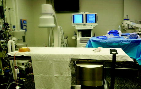
Fig. 37.2
Fluroscopy table. The fluoroscopy table can be imaged through and also has sufficient clearance to allow the C-arm to move freely. PICTURE COURTESY OF: Colleen F. DeCarlis, RN, BS
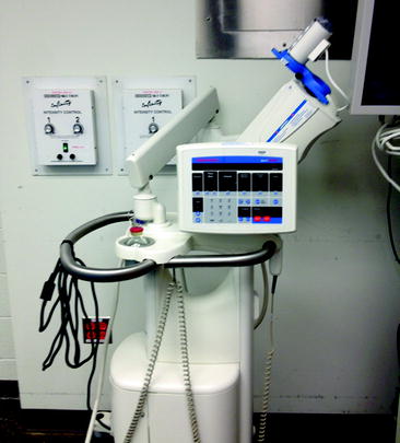
Fig. 37.3
Power injector. The power injector allows for improved image quality. PICTURE COURTESY OF: Colleen F. DeCarlis, RN, BS
Space considerations also relate to radiation safety in that the level of radiation is reduced by the square of the distance to the source (inverse square law). As such, as the endovascular suite becomes larger, they become safer and more comfortable, but at the expense of mobility and maximizing the resource of space. The repeated use of fluoroscopy requires the endovascular team to monitor potentially unsafe levels of radiation. This risk is reduced by monitoring radiation levels, wearing protective shielding, and minimizing fluoroscopy time. In the civilian hospital, great care is also taken to verify that radiation levels outside the suite are negligible. Patient safety should not be overlooked in the development of the endovascular suite. Anesthesia is usually intravenous sedation with local supplementation. Cardiopulmonary monitoring is performed by an anesthesiologist, CRNA, or nurse using continuous electrocardiogram and intermittent blood pressure measurements.
3 Disease Distribution
In 2005, Fox et al published the first report of the distribution of vascular injuries seen in the current conflicts in Iraq and Afghanistan. From December 2001 through March 2004, all wartime evacuees evaluated at a single institution were prospectively entered into a database and retrospectively reviewed. Of 3,057 soldiers evacuated for medical evaluation, 1,524 (50%) sustained battle injuries. Known or suspected vascular injuries occurred in 107 (7%) patients, and these patients comprised the study group. Sixty-eight (64%) patients were wounded by explosive devices, 27 (25%) were wounded by gunshots, and 12 (11%) experienced blunt traumatic injury. The majority of injuries (59/66 [88%]) occurred in the extremities. Nearly half (48/107) of the patients underwent vascular repair in a forward hospital in Iraq or Afghanistan. Twenty-eight (26%) required additional operative intervention on arrival in the USA. Vascular injuries were associated with bony fracture in 37% of soldiers. Twenty-one of the 107 had a primary amputation performed before evacuation. Amputation after vascular repair occurred in eight patients. Of those, five had mangled extremities associated with contaminated wounds and infected grafts. Sixty-seven (63%) patients underwent diagnostic angiography. The most common indication was mechanism of injury (42%), followed by abnormal examination (33%), operative planning (18%), or evaluation of a repair (7%). The conclusions of this study were that wounding patterns reflected past experience with a high percentage of extremity injuries. Management of arterial repair with autologous vein graft remains the treatment of choice. Repairs in contaminated wound beds should be avoided. An increase in injuries from improvised explosive devices in modern conflict warrants the more liberal application of contrast arteriography. Endovascular techniques have advanced the contemporary management and proved valuable in the treatment of select wartime vascular injuries. This was confirmed in a later report by Clouse et al when they reported on their series of 408 arterial injuries managed in theater.
In 2006, Weber et al published a larger series on the modern military trauma experience with upper extremity trauma. In this report of 58 patients with upper extremity arterial wounds there were a total of 63 distinct arterial injuries. The axillary artery was injured in eight patients (13%). The brachial artery was injured in 33 patients (52%). The radial artery was injured in 16 patients (25%), and the ulnar artery was injured in six patients (10%).
The modern management of venous injuries suffered during military trauma was reported by Quan et al in 2006. In this report or 81 venous injuries in 65 patients, 43 (66%) patients suffered concomitant arterial injuries and 11 (17%) multiple venous injuries. Venous injury from improvised explosive devices was seen in 44 (67.7%) patients, gunshot wounds in 18 (27.7%), and motor vehicle accidents in 3 (4.6%). Extremity injuries accounted for a large number of wounds, comprising 64.2% of the cases, with 19.7% torso and 16.1% neck wounds. Ligation was the most common modality of treatment in combat zones. In a later report on venous injuries, Quan reported to find no difference in the incidence of venous thromboembolic complications between venous injuries managed by open repair versus ligation. Venous injuries were treated by ligation in 65 patients (63.1%) and by open surgical repair in 38 (36.9%). Postoperative extremity edema occurred in all patients irrespective of method of management. Thrombosis after venous repair occurred in 6 of the 38 cases (15.8%). Pulmonary emboli developed in three patients, one after open repair and two after ligation (P > 0.99).
4 Diagnosis
The diagnosis of vascular injuries still depends greatly on physical exam findings. Previous studies have shown little value in performing arteriography for proximity as a sole indication in civilian trauma. In general, performing ankle brachial indices can select out those patients at high risk for vascular injury that need arteriography. Patients with an ABI <0.9 has been shown to correlate with a higher incidence of vascular injury in civilian trauma patients. In contrast, occult vascular injuries requiring further intervention occur at a much higher frequency in combat. As such, liberal imaging is advocated, even in patients with a diffuse injury pattern and a normal physical exam. Recent combat casualties have stimulated a reassessment of the principles of management of high-risk extremity injuries with a normal vascular examination. Rapid evacuations have presented numerous US soldiers to the USA for evaluation in the early postinjury period. The liberal application of arteriography is a low-risk method to provide high-yield data in the delayed vascular evaluation of extremities injured from modern military munitions. Physical examination findings remain the most useful indicator, but a normal examination can be misleading and should not guide the decision for invasive imaging. Lesions are found and require further intervention at a higher rate than expected from the typical civilian trauma experience.
5 Diagnostic Imaging: Ultrasound/CT/MRI/Diagnostic Angiography
The use of handheld Doppler during Vietnam as reported by Lavenson et al sets the standard for the field assessment of vascular trauma patients. Today duplex ultrasonography has extended the noninvasive diagnostic capabilities of trauma surgeons. The focused abdominal scan (FAS) has become the standard of care in both civilian and military trauma to date. Over time duplex ultrasonography has become easily portable and can be applied rapidly in most field hospital situations. For these reasons, ultrasound is well suited as an adjunct to physical exam in diagnosing vascular injury. However, its usefulness in the assessment of extremity vascular injuries can be limited. As in any situation, the accuracy of ultrasound is user dependent in diagnosing vascular injury.
Multislice (2–16) computed tomography (CT) (Fig. 37.4) is a technique that is now applied to many military combat support hospitals. It is currently designed to be deployed in its own iso-shelter as an adjunctive component. The usefulness of these low slice CT scans is, however, limited by their slow speed and the fact that metal artifact is created by bullet fragments and shrapnel commonly found in war military trauma. Newer generation 64 slice CT angiography, however, has been shown to be reliable in the diagnosis of occult vascular injury, even in the presence of bullet fragments, shrapnel and orthopedic hardware. CT also has the advantage that the entire arterial system can be imaged with a single, well-timed contrast bolus. This modality is quickly becoming the imaging method of choice in military evacuation and rear echelon hospitals.
Fig. 37.4


Related posts:


Stay updated, free articles. Join our Telegram channel
Full access? Get Clinical Tree
