Fig. 31.1
The saphenous eye of the great saphenous vein. Transverse B mode ultrasound image of GSV (a). Probe position on the thigh (b)
(c)
The great saphenous vein (GSV) starts in the medial foot, ascends in the leg in the saphenous sheath anterior to the medial malleolus, and runs along the medial border of the tibia. It passes medial to the knee and then runs medial to the femoral vein. At the groin, it drains into the common femoral vein at the saphenofemoral junction (SFJ).
(d)
Multiple perforator veins perforate the superficial fascia and route blood from superficial to deep veins.
2.
Some individuals have accessory saphenous veins, which can also run in sheaths with ultrasound saphenous eyes. The anterior accessory saphenous vein (AASV), often present, runs lateral to the GSV. The AASV can be distinguished from the GSV by the alignment sign, or its position superficial to the femoral vein and artery on transverse ultrasound.
3.
The SFJ has a constant terminal valve located 1–2 mm distal to the SFJ on the GSV, and usually another preterminal valve 2 cm distally along the GSV, marking the limit of the SFJ. Proximal veins draining the abdominal wall and pudendum terminate at the SFJ. Additionally, distal veins, such as the AASV or posterior accessory saphenous vein, often drain at or near the SFJ.
4.
The small saphenous vein (SSV) begins in the lateral foot, ascends in the leg posterior to the lateral malleolus, then runs in the midline posterior calf between the two heads of the gastrocnemius to the popliteal fossa. The SSV, like all saphenous veins, runs in a sheath with the ultrasound saphenous sign. It terminates most often at the SPJ, draining into the popliteal vein superior to the popliteal crease. The SSV often continues as a thigh extension draining into deep veins more proximally, or can also continue as the Giacomini vein, draining into the GSV.
2 Disease Definition
Patients have chronic venous disease when they have either symptoms (pain or swelling) or physical signs (varicose veins, telangiectasias, dermatologic complications) attributable to abnormal venous flow or venous reflux.
(a)
Varicose veins are blue dilated subcutaneous veins usually over 3 mm diameter, and they most often bulge outside the skin surface.
(b)
Reticular varicosities are blue dilated subdermal veins usually less than 3 mm in diameter.
(c)
Telangiectasias, also called spider veins, are red or purple dilated intradermal venules usually less the 1 mm in diameter.
(d)
Chronic venous insufficiency is diagnosed when a patient has either edema or dermatologic complications from chronic venous disease.
(e)
Dermatologic complications of venous reflux: An ulcer is a full thickness skin defect. Lipodermatosclerosis is a localized fibrosis of the skin and subcutaneous tissues. Atrophie blanche is a localized area of white, atrophic skin areas surrounded by dilated capillaries. Venous pigmentation is brown and due to extravasated blood. Venous eczema is red and can weep, blister, or scale. Corona phlebectactica is a fan-shaped pattern of small intradermal veins.
3 Disease Distribution
Chronic venous disease signs, such as telangiectasias, varicose veins, and venous ulcers, are common but notoriously underdiagnosed. Telangiectasias have over 80% prevalence, while varicose veins have around 25% prevalence. More severe vein disease is thankfully less common, but around 1% of the population has an active or healed venous ulcer.
A first-degree family history of varicose veins in a parent is the greatest risk factor for the development of chronic venous disease, with a corrected odds ratio of over 2. Other risk factors are increased age, female sex, pregnancy, high body mass index, taller height, and prolonged standing.
4 Disease Classification
(a)
Chronic venous disease is classified using the CEAP system, based on clinical manifestations (C), etiology (E), anatomic disease distribution (A), and pathophysiology (P). The “C” from CEAP is most often used in venous studies and in clinical practice.
(b)
C6 patients have an active venous ulcer.
(c)
C5 patients have a healed venous ulcer.
(d)
C4 patients have dermatologic complications from venous disease other than ulcer, as discussed in IId.
(e)
C3 patients have venous edema.
(f)
C2 patients have varicose veins.
(g)
C1 patients have telangiectasias and/or reticular varicosities.
(h)
C0 patients have no visible signs of venous disease. These patients can still have pain or swelling from venous reflux.
5 Diagnosis: Clinical
(a)
Pain is the most common symptom. Patients often describe their pain as aching, heaviness, fatigue, soreness, or burning. Location is typically at varicose vein sites, or along the path of the GSV (medial ankle, calf, and/or thigh) or SSV (posterior or distal lateral calf), but can also consist of generalized leg pain. It usually worsens with prolonged standing and improves with extremity elevation.
(b)
Swelling is also common. Venous edema usually begins around the medial malleolus, although it can spread proximally throughout the extremity. It typically worsens with prolonged standing and improves with extremity elevation.
(c)
Telangiectasias, varicose veins, swelling, and skin changes are the common signs.
6 Diagnostic Imaging
Duplex ultrasound is the key to diagnosis and management of chronic venous disease. This test rules out deep venous thrombosis as a cause of patient symptoms. It also defines the anatomy and extent of venous reflux critical in determining patient management. It is important to note that most ultrasound studies performed on the lower extremity evaluate only the presence or the absence of thrombus and do not investigate the patient’s valvular function. Therefore, a “negative” venous ultrasound usually does not rule out the possibility of valvular insufficiency unless it is performed in a vascular lab that specializes in venous disorders. In phlebological ultrasound, superficial, deep, and perforator systems are examined for reflux and obstruction. Superficial venous examination is performed along the course of all saphenous veins and their junctions, as well as any other refluxing tributary veins. The source of reflux for all superficial varices should be determined.
Venous reflux is defined as blood flow in the reverse direction to physiologic flow (for example, towards the floor in a standing patient). Pathologic reflux lasts for more than 0.5 s. Reflux can be elicited by several methods including calf squeeze, muscle squeeze, vein cluster manual compression, Valsalva maneuver, active foot dorsiflexion and relaxation, or pneumatic calf cuff deflation. The most common finding in patients with varicose veins is reflux within the GSV.
Occasionally, patients may need magnetic resonance venography, if ultrasound is insufficient to fully evaluate the relevant pathology. Patients with unusual skin or duplex ultrasound findings may have a vascular malformation. Iliac vein compression can present with severe pain and swelling which can be posttraumatic or congenital (May-Thurner Syndrome, in which the left iliac vein is compressed by the right iliac artery). Patients can have pelvic congestion syndrome, with pelvic pain and varicosities due to pelvic vein reflux. This can be confirmed by venography.
7 Management
Table 31.1 lists the indications for treatment of superficial venous reflux. The main contraindication for superficial venous ablation is superficial or deep venous obstruction.
Table 31.1
Indications for superficial venous treatment
General appearance |
Significant pain attributable to venous disease |
Significant edema attributable to venous disease |
Skin changes attributable to venous disease |
Leg ulcer attributable to venous disease |
History of superficial thrombophlebitis |
History of bleeding varicose veins |
Compression therapy is used for patients with any level of venous reflux. Compression improves venous hemodynamics, improves quality of life for patients with chronic venous disease, reduces edema and skin discoloration, and improves the venous ulcer-healing rate. The main contraindication to compression use is peripheral arterial disease, especially with ankle-brachial index less than 0.5. Commercially available compression stockings are most often used. Inelastic compression, such as Unna boots and multilayer compression dressings are also used in patients with active ulcers.
Refluxing veins are corrected in the following order: great saphenous, small saphenous, other superficial veins, perforator vein(s), deep vein(s). The main contraindication to reflux treatment is venous obstruction. Indications for perforator vein ablation are controversial, but it is generally accepted that correction of perforator reflux is indicated for patients with class C5–C6 disease. In addition, perforator reflux should be reevaluated by duplex ultrasound following successful superficial ablation because they often normalize afterward, especially if the patient has no deep venous reflux. Deep venous reflux treatments carry a higher risk, have more variable success rates, and are usually treated only for patients with particularly severe symptoms, and only at specialized centers.
8 Open Operative Choices
(a)
The classic surgical procedure for GSV reflux is ligation of the GSV at the SFJ and saphenous trunk stripping. Often the below-knee GSV is preserved due to a significant risk of saphenous nerve injury. This surgical procedure has become less common, as the risks for recurrent varicosities after treatment due to neovascularization have become clear. SSV surgery is also possible, but is plagued by highly variable anatomy, high varicosity recurrence rates, and injuries to the popliteal vein, popliteal nerve, and sural nerve.
(b)
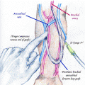
Ambulatory phlebectomy (AP) can be used for remaining visible and palpable tributary or localized varicosities after superficial axial vein ablation. In this method, the patient’s varicosities are marked preoperatively in the standing position. The patient is placed supine and tumescent anesthesia is administered. One to 3 mm incisions are made vertical to the varicosity, which is hooked and then grasped by a hemostat or a Size 3 Oesch hook. Gentle traction is then applied to tease the vein out of the wound. The vein is divided and the ends extracted by a combination of rotation, traction, and massage. At the completion of the procedure, a compression dressing is applied and the patient is ambulated. Complications are rare but include blister formation, transient pigmentation, and telangiectatic matting, with superficial thrombophlebitis, hematoma, and dysesthesias less common.
(c)
Transilluminated, powered phlebectomy


Related posts:

Stay updated, free articles. Join our Telegram channel
Full access? Get Clinical Tree
