Chapter 189
Arteriovenous Malformations
Epidemiology
Arteriovenous malformations (AVMs) are developmental malformations of the vascular system that result in abnormal communication between arteries and veins. The primary abnormality appears to be at the level of the capillary bed. AVMs are classified as a type of vascular malformation using the classification system of Mulliken and Glowacki. AVMs also include arterial malformations and arteriovenous fistulae and are characterized as high flow lesions.
Clinical Findings
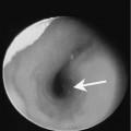
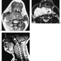
The clinical presentation can be variable depending on the extent of the lesion. The neck and craniofacial region are common sites of occurrence. On clinical examination, AVMs present as a soft tissue fullness that is compressible. Superficial AVMs may be associated with a palpable thrill or audible bruit. The overlying skin may be discolored due to dilated superficial veins. Other presentations in more advanced lesions include facial deformity, skin ulceration, or functional compromise. Patients may also present with hemorrhage that may be spontaneous or may follow minor trauma such as tooth extraction. AVMs may enlarge with pregnancy. It is unclear as to whether the growth is due to direct hormonal stimulation or whether it results secondarily from an increase in the circulating blood volume.
Pathology
AVMs result from abnormal development of the arterial, capillary, and venous components of the vascular system. The lesions grow commensurately with the individual and show no evidence of endothelial proliferation. The vascular channels are lined by mature endothelium with normal mitotic activity.
Treatment