Abstract
All imaging modalities may be compromised by artifacts that obscure malignancy or mimic malignancy. FDG PET/CT may be compromised by a multitude of artifacts, primarily associated with technical performance of PET/CT scanners and unexpected biodistribution of FDG. A few of the more common artifacts on FDG PET/CT are discussed in this chapter, including motion, attenuation correction, partial volume effects, suboptimal FDG biodistribution, and blooming artifacts.
Keywords
FDG, PET/CT, artifacts, motion, attenuation correction, partial volume effects, blooming artifact
All imaging modalities may be compromised by artifacts which obscure malignancy or mimic malignancy. 18F-fluorodeoxyglucose positron emission tomography/computed tomography (FDG PET/CT) may be compromised by a multitude of artifacts, primarily associated with technical performance of PET/CT scanners and unexpected biodistribution of FDG. A few of the more common artifacts on FDG PET/CT are discussed here.
Motion
Patient motion during acquisition of PET/CT images may introduce multiple artifacts which need to be recognized. The PET/CT scanner is composed of separate PET and CT imaging elements. Thus, although we refer to PET/CT images, it is important to remember that separate PET and CT images are obtained and then fused together. Under idealized conditions we expect there to be perfect alignment of the PET and CT images; however, this is virtually never the case. Motion of the patient between acquisition of the CT images and the PET images will result in misregistration of the combined PET/CT images, which may obscure an otherwise appreciable malignancy. This is common in the head and neck, if the head is not immobilized. This will also produce incorrect attenuation correction maps from the CT images and lead to incorrect calculation of standardized uptake values (SUVs).
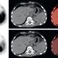
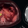
Even if voluntary muscular motion of the head/neck and extremities is perfectly controlled for, involuntary motion will invariably affect PET/CT images. Motion of the diaphragm will cause misregistration between CT and PET images in the lower lungs and upper abdomen, sometimes quite significantly. This could result in artifactual placement of liver lesions over the lung on PET ( Fig. 23.1 ), or vice versa. Incorrect localization of photons and suboptimal attenuation correction maps from diaphragmatic motion will artifactually lower SUVs for lesions near the diaphragm. In the extreme, this problem may cause lesions in the lower lungs or upper abdomen to artifactually disappear from PET images ( Fig. 23.2 ). Careful evaluation of the CT images near the diaphragm is important to prevent these errors.


Related posts:


Stay updated, free articles. Join our Telegram channel

Full access? Get Clinical Tree


