Abstract
The brain demonstrates substantial physiologic FDG avidity. Malignancy involving the brain, including metastases, lymphoma, and primary gliomas, may be detected either as foci of FDG avidity greater than physiologic brain background, areas of FDG photopenia, or as lesions on the corresponding CT scan. FDG PET/CT plays an important role in distinguishing recurrent malignancy from benign radiation necrosis following radiation therapy of brain malignancy. Benign findings resulting from seizures and infarcts must be distinguished from brain malignancy.
Keywords
FDG, PET/CT, brain, brain cancer, radiation necrosis, lymphoma, infarcts, seizures
Normal Brain
18F-Fluorodeoxyglucose (FDG) was initially designed to be a brain metabolism tracer and today has multiple applications in neurologic disorders, such as dementias and seizures. The demonstration of the value of FDG in metabolically active tumors has led to a vast majority of clinical FDG positron emission tomography/computed tomography (PET/CT) studies being performed for evaluation of malignancy, and this chapter will focus on FDG PET/CT for oncologic findings in the brain.
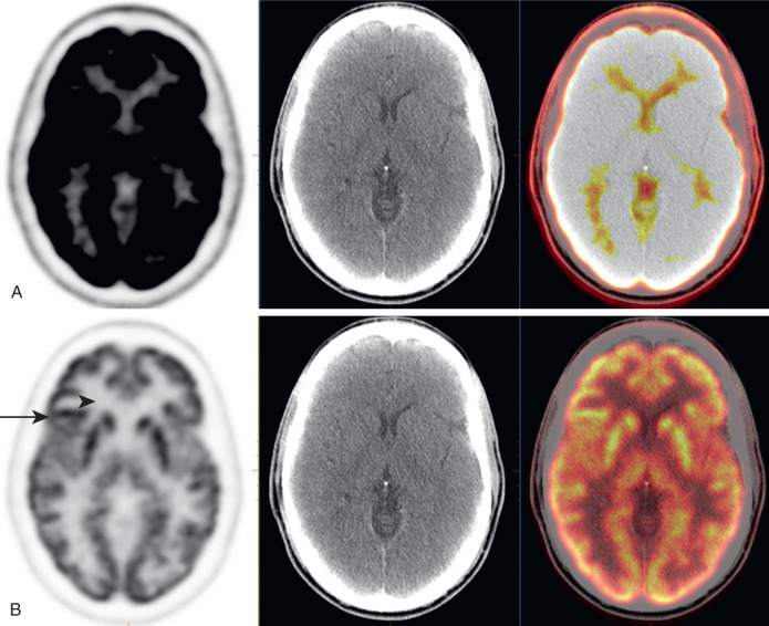
The brain accounts for about 20% of the body’s glucose consumption; thus the brain demonstrates very high physiologic uptake on FDG PET. To view the brain appropriately, it will be necessary to optimize the PET window settings. For example, when the PET window is set at a maximum standardized uptake value (SUV) display of 5, all voxels with an SUV of 5 or above will appear identically black on a gray-scale display. This is a common setting for evaluating most of the body, but the brain will appear nearly uniformly black and provide little information. By changing the PET window to a maximum SUV display of 15, 20, or more, the details of the brain will become visible ( Fig. 6.1 ). Each brain you examine may have a different PET window for optimal evaluation.
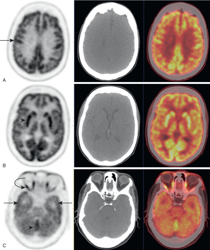
Once the display is properly windowed for the brain, the metabolism of the brain becomes evident ( Fig. 6.2 ). Brain FDG avidity is normally symmetric, higher in the gray matter than white matter. Gray matter gyri will be apparent. There may be slightly lower uptake in the temporal lobes than the frontal, parietal, and occipital lobes. The basal ganglia often have the highest intracranial physiologic uptake. There may also be slightly higher uptake in the midline cerebellum. Extracranially, the optic muscle may be highly FDG avid.
Initial Detection of Brain Malignancy
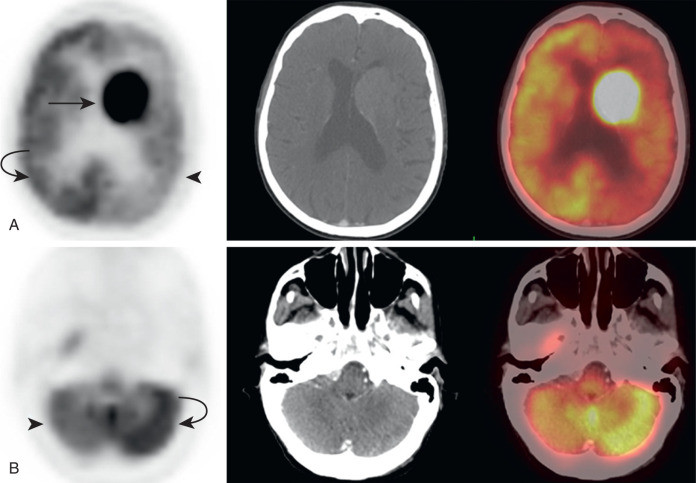
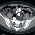
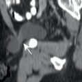
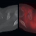
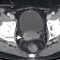
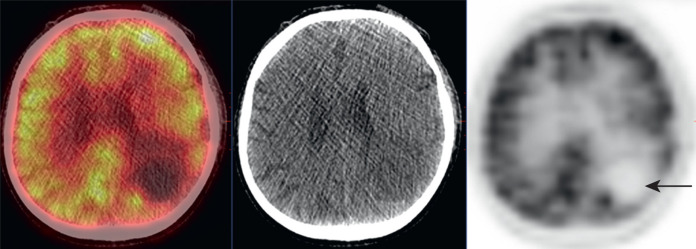
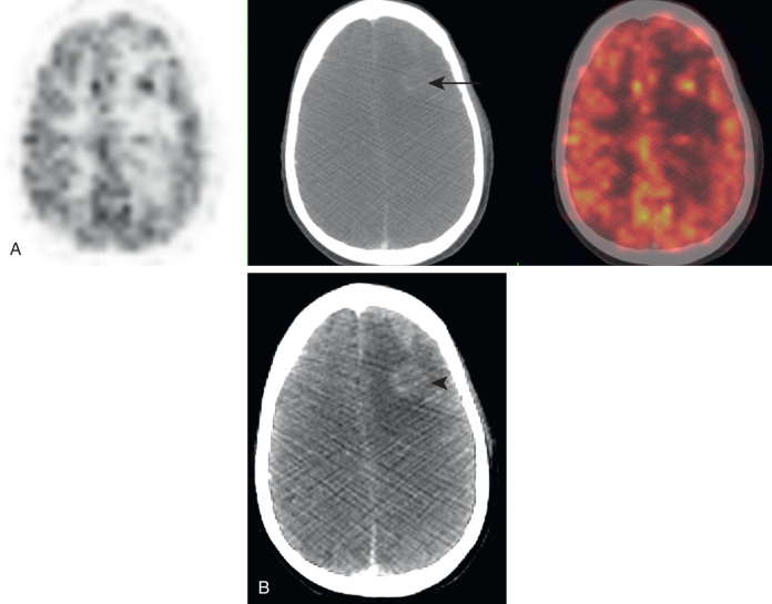
Malignancy involving the brain includes metastases, lymphoma, and primary brain gliomas. Due to the high physiologic FDG uptake in normal brain, visualization of FDG-avid intracranial malignancies may be obscured. High-grade metastases may exhibit FDG avidity even greater than brain background and be identified as foci of high FDG avidity ( Fig. 6.3 ). The corresponding anatomic mass may not be visualized on noncontrast CT, or even contrast-enhanced CT. Contrast-enhanced multiparametric magnetic resonance (MR) is the most sensitive method for the initial detection of brain metastases (see Fig. 6.3B ). However, not all brain metastases will have FDG avidity greater than the normal brain. In cases where FDG avidity is less than normal brain, metastases may be visualized as photopenic defects in normal brain avidity ( Fig. 6.4 ). In some cases, the avidity of metastases may not be discernible from normal brain avidity. In these cases, evaluation of the corresponding CT images may reveal metastases that are not apparent on FDG PET ( Fig. 6.5 ). In all cases, inconclusive findings can be further evaluated by contrast-enhanced multiparametric MR.
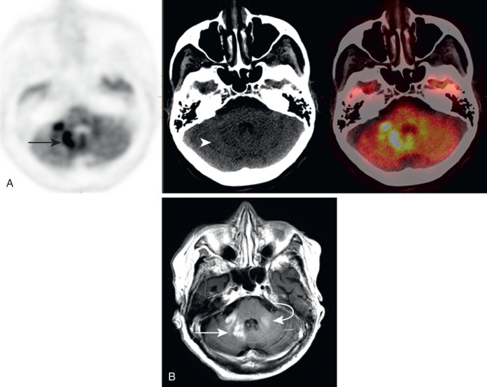
High-grade lymphomas, particularly primary central nervous system (PCNS) lymphomas, will usually be more FDG avid than normal brain and discernible as FDG-avid foci ( Fig. 6.6 ). Lower-grade lymphomas may not be apparent on FDG PET. Again, MR will be the most sensitive method for initial detection of disease extent in the brain. A specific indication for brain FDG PET/CT is in acquired immunodeficiency syndrome (AIDS) patients with a new enhancing lesion on MR. The most common differential diagnoses for this scenario are toxoplasmosis and AIDS-related lymphoma. An FDG PET/CT may assist in making this distinction, as AIDS-related lymphoma is normally more FDG avid than background brain, while toxoplasmosis is relatively photopenic ( Fig. 6.7 ).