Abstract
Optimal FDG PET/CT relies upon proper performance of the examination and reporting of the results. This chapter discusses proper patient preparation for FDG PET/CT, quantification of FDG uptake, and structured reporting imaging findings.
Keywords
FDG, PET/CT, SUV, structuring reporting
Performing FDG PET/CT
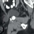
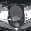
Patients need to be prepared for 18F-flourodeoxyglucose (FDG) positron emission tomography/computed tomography (PET/CT) in order to optimize the distribution of FDG in the body. Patients should avoid exercise starting the day before the examination in order to prevent muscular FDG uptake. Patients should not consume food or liquids other than unflavored water for at least 4 hours prior to administration of FDG, in order to prevent insulinemia and FDG uptake in muscles and fat. Hydration with a liter of water in the hours before the scan will help dilute excreted FDG in the urine, both reducing patient radiation exposure and artifacts. The patient should be kept warm, starting before FDG administration, in order to prevent FDG-avid brown fat. Blood glucose level is measured before FDG administration, as a glucose level greater than 200 mg/dL may alter FDG biodistribution. FDG is administered intravenously with the patient sitting or lying comfortably, and the patient should remain in this position to minimize muscular uptake. If possible, the patient should remain silent to prevent FDG uptake in vocal musculature, particularly for patients with cancer in the head/neck. The patient should void before being positioned on the PET/CT scanner to remove excreted FDG in the urine. PET/CT imaging usually begins about 60 minutes after FDG administration. The extent of FDG uptake in tissues changes with time; thus it is important to keep the uptake times relatively similar in order to best compare studies. The patient is positioned comfortably on a PET/CT scanner for the examination, which may take 30 to 60 minutes, depending on the field of view and number of minutes per bed position. The typical field of view is the mid-skull to mid-thigh. Additional views of the neck, brain, or extremities may be obtained in selected patients. See Box 2.1 for a list of these maneuvers. Following imaging, CT images are reconstructed and used to create an attenuation map in order to attenuation correct the PET images. CT, nonattenuation corrected PET, and attenuation corrected PET images are produced. The attenuation corrected PET images are usually fused to the CT images to allow evaluation of fused PET/CT images. All images are displayed in multiplanar reconstructions.
- 1.
Avoid exercise starting the day before the FDG PET/CT.
- 2.
No food or liquids other than water starting at least 4 hours before the FDG PET/CT.
- 3.
Hydration with a liter of water in the hours before FDG administration.
- 4.
Keep patient warm starting before FDG administration.
- 5.
Measure blood glucose level before FDG administration.
- 6.
Administer FDG intravenously while patient sits or lies comfortably and quietly.
- 7.
Patient should void before being placed on PET/CT scanner.
- 8.
Position patient comfortably on scanner for the relatively long image acquisition. Imaging begins about 60 minutes following FDG administration.
Quantifying FDG Avidity With Standardized Uptake Values
FDG avidity is increasingly being quantitated, in order to have numerical values for comparisons. Quantitation of FDG avidity is most commonly performed with standardized uptake values (SUV), a measurement of tracer uptake in a lesion normalized to a distribution volume. There are many different formulas of SUV, depending on how the SUV is normalized (weight, lean body mass [LBM], body surface area), and how the region of interest (ROI) is analyzed (SUVmax, SUVmean, SUVpeak, etc.). Most commonly, SUVmax is reported, which is the SUV normalized for body weight for the most FDG-avid voxel within a ROI. SUV normalized for LBM is called SUL, as is recommended by several organizations. SUL provides less variation in the calculated value than SUV normalized for body weight. However, SUVmax is so highly reproducible and easy to calculate that it is currently the most commonly utilized measure of FDG avidity in clinical practice. SUVmax is calculated as SUVmax = tracer uptake in ROI / (injected activity / patient weight). This provides a number (2, 10, 20, etc.) that conveys a sense of the relative extent of FDG uptake in a lesion.
It is important to remember that calculated SUV values are “semiquantitative.” If you perform an FDG PET/CT in the same patient on two consecutive days, you may get slightly different SUV values, despite the lesion truly being the same. This is because the amount of FDG uptake in a lesion is dependent on multiple biologic and technical factors ( Box 2.2 ). For example, higher blood glucose levels may reduce FDG uptake in tumors and a long FDG uptake time (time between FDG injection and image acquisition) often results in increased FDG uptake. To minimize variations in SUV because of these biologic and technical factors, a number of guidelines are suggested for the performance on FDG PET/CT ( Box 2.3 ). These biologic and technical factors often result in a 10% to 20% difference in SUV, even if there is no change in the tumor.
- A.
Biologic factors
- 1.
Patient weight
- 2.
Blood glucose level
- 3.
Insulin
- 4.
FDG uptake time
- 5.
Lesion size (partial volume effects, see Chapter 23 )
- 1.
- B.
Technical factors
- 1.
Variability in different scanners
- 2.
Variation in calibration of scanners
- 3.
Variability in reconstruction methods
- 4.
Motion
- 1.
Related posts:


Stay updated, free articles. Join our Telegram channel

Full access? Get Clinical Tree


