Clinical Presentation
The patient is an 86-year-old man with prostate carcinoma who presented with history of several months of progressive right upper neck pain and recent development of suboccipital headaches. No pain or numbness reported in the upper extremities. Limited range of motion was observed in the right upper extremity. Otherwise, the physical examination was normal.
Imaging Presentation
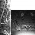
Radionuclide bone scan reveals focal area of increased activity in right upper cervical spine region ( Fig. 33-1 ) . Otherwise the bone scan is normal. Magnetic resonance (MR) imaging obtained both without and with intravenous contrast enhancement reveals abnormal T1 and T2 signal intensity in and adjacent to the right C1-2 facets. Contrast enhancement is demonstrated in the C1-2 facets and adjacent bone marrow, in the periarticular soft tissues, and within the right C1-2 facet joint ( Figs. 33-2 to 33-4 ) . Findings indicate active right C1-2 inflammatory facet arthropathy (facet synovitis).


Discussion
Osteoarthritis commonly occurs in the cervical zygapophyseal (facet) joints in patients with advancing age. Any cervical level may be affected by facet osteoarthritis. Osteoarthritis has been reported to involve the atlantoaxial (C1-2) facet joints (also called the lateral atlantoaxial joints ) in approximately 4% of patients overall, and the prevalence increases significantly after the fifth decade of life.
Osteoarthritis involving the C1-2 facet joints can produce a distinct clinical syndrome characterized by limitation of neck rotation, severe upper neck pain, and occipital headaches. The patient may have suboccipital pain trigger points, suboccipital crepitus upon palpation, and a rotational head tilt deformity may be present. Upper neck pain and occipital neuralgia can also be secondary to osteoarthritis involving the atlantal-odontoid joint (anterior C1-2 joint). Lateral and anterior C1-2 osteoarthritis commonly coexist (see Figs. 33-3 and 33-4 ).
The joints between the lateral masses of C1 and C2 anatomically are considered to be zygapophyseal (facet) joints. These joints have some unique anatomic features that are not present in the lower cervical facet joints. The inferior C1 (atlas) articular facet is relatively flat, and this articulates with the relatively convex C2 (axis) superior articular facet. This incongruent configuration of the C1-2 articular facets results in a rather wide (3 to 5 mm) atlantoaxial facet joint space anteriorly and posteriorly. The shape of these joints and the relatively loose capsule that normally surrounds these joints imparts greater joint mobility to these joints than is possible at other cervical levels. The greatest degree of neck rotation occurs at this level.
Meniscus-like synovial folds are present within the C1-2 facet joints during infancy, but these folds usually involute and are not present in older children and young adults. As the atlantoaxial joints age or degenerate, meniscus-like synovial folds again develop that fill the incongruous spaces within the C1-2 joints. These folds are believed to provide stability to these joints. Such meniscal folds also develop in degenerated facet joints elsewhere in the spine.
The earliest findings in degeneration of the lateral atlantoaxial joints is superficial flaking of the articular cartilage. Thinning and fibrillation of the articular cartilage with associated facet cortical sclerosis and irregularity also occur as in any other degenerating facet joint. Eventually the joint space narrows and hypertrophic bone arises at the bone margins of the joint ( Figs. 33-5 to 33-8 ) .


