ATRIOVENTRICULAR
CONNECTION, DOUBLE INLET
VENTRICLE, AND TRICUSPID
ATRESIA WITH
VENTRICULAR SEPTAL
DEFECT
16
 UNIVENTRICULAR ATRIOVENTRICULAR CONNECTION
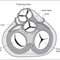
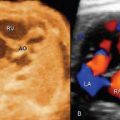
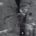
Univentricular atrioventricular connection describes a group of cardiac malformations where the atrioventricular connection is completely or predominantly to a single ventricular chamber. Embryologically, this malformation is thought to result from failure of the development of the bulboventricular loop stage. Much debate still exists today on the various subclassifications of cardiac anomalies within this group and what should be included or excluded. From a clinical point of view, a congenital heart defect with a univentricular atrioventricular connection, single ventricular physiology, describes a heart with one functioning ventricle with inflow from one or both atria. Numerous terms were used to describe this malformation, including univentricular heart, primitive ventricle, common ventricle, single ventricle, cor triloculare biatriatum, cor biloculare, dominant ventricle, and double inlet ventricle (1). The classic Van Praagh’s classification (2), which was later modified by Hallermann et al. (3), described one or two atrioventricular valves that empty into a single ventricle and excluded mitral or tricuspid atresia. Anderson’s simpler classification described a single ventricular mass with or without a rudimentary chamber and allowed for the inclusion of mitral or tricuspid atresia (4,5). In Anderson’s classification, the rudimentary chamber, if present, should not have an inlet but may have an outlet (4,5). Within univentricular atrioventricular connection, three subgroups can be identified: double inlet, where two atria connect to a single ventricle through two patent atrioventricular valves; single inlet, where one atrium connects to a single ventricle through a single atrioventricular valve; and common inlet, where both atria connect to a single ventricle through a single atrioventricular valve (6). The morphology of the ventricle is generally a left ventricular morphology with a rudimentary right chamber. On rare occasions, a right ventricular morphology with a rudimentary left chamber, or a ventricle of indeterminate morphology without a rudimentary chamber, can be seen. A single ventricle heart, which results from a surgical repair of a congenital heart anomaly, should not be classified as univentricular atrioventricular connection. Table 16-1 lists several cardiac anomalies that may show a single ventricle on fetal echocardiography. Of those, double inlet ventricle and tricuspid atresia with ventricular septal defect have been commonly classified in the univentricular atrioventricular connection and will be discussed in this chapter. Figure 16-1 represents four-chamber views in two fetuses with single ventricle anatomy.
 DOUBLE INLET VENTRICLE
Definition, Spectrum of Disease, and Incidence
Double inlet ventricle (DIV) is considered a classic and most common form of univentricular atrioventricular connection (6). It is characterized by two normally developed right and left
Related posts:

Stay updated, free articles. Join our Telegram channel

Full access? Get Clinical Tree


