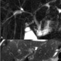
85 Avascular Necrosis of the Hip Pelvic and hip MRI is typically obtained utilizing a body coil, enabling simultaneous bilateral imaging. Unilateral imaging with a dedicated surface coil, however, allows increased SNR and spatial resolution. The detection and treatment of early avascular necrosis (AVN) of the femoral head—the most common location for this condition in the body—is the primary indication for hip MRI. SI characteristics of femoral head AVN correspond well with underlying pathology: the commonly described double-line sign within the femoral head refers to the appearance of a hyperintense line immediately adjacent to linear hypointense signal on T2WI or PDWI. The former line correlates pathologically with granulation tissue and the latter with reactive sclerotic bone and fibrosis. Together the double line represents the interface between normal and necrotic marrow. The Mitchell system classifies the MRI progression of femoral head ischemia. Early, lesions (class A) demonstrate fat-like SI as shown in the left femoral head of Figs. 85.1A,B. Here, (A) T1WI demonstrates isointensity of the lesion to normal marrow superior to a hypointense line correlating with a nidus of fibrovascular proliferation and demarcating pathologic from normal bone. A full double-line sign is not seen, as is the case 20% of the time. On (B
![]()
Stay updated, free articles. Join our Telegram channel


Full access? Get Clinical Tree


