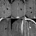
18 Multiple Sclerosis An international committee has developed specific MRI criteria for the diagnosis of MS, which include three of the following disseminated in time: (1) a gadolinium-enhancing brain or spinal cord lesion or a total of nine T2-hyperintense lesions, (2) one infratentorial or spinal cord lesion, (3) one juxtacortical lesion, and (4) three periventricular lesions. MRI is the most sensitive test for MS, although findings may be confused with those of small vessel ischemia. A younger, female patient favors MS, and given the high prevalence of small vessel disease, older patients must not be diagnosed with MS based solely on imaging. Figure 18.1A demonstrates the typical pattern of disease on FLAIR scans, with small focal areas of abnormal high SI—correlating pathologically with edema and gliosis—scattered throughout the periventricular white matter. Periventricular lesions are nonspecific for MS, as opposed to callosal lesions— best seen on sagittal FLAIR images as in Fig. 18.1B (white arrow). These are typically oval-shaped with a flat inferior border, line the ependymal surface, and are oriented perpendicular to the lateral ventricle. Such findings are sometimes overlooked on axial scans (Fig. 18.1A, black arrow
![]()
Stay updated, free articles. Join our Telegram channel


Full access? Get Clinical Tree


