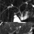
42 Common Incidental Findings Benign vertebral body hemangiomas are a common incidental finding in the spine. Larger lesions may bleed, expand to compress the spinal canal, or weaken the vertebral body leading to fracture, although all of these complications are very rare. Hemangiomas consist of adipose and angiomatous tissue. As illustrated in Fig. 42.1, the fat within these lesions results in a high SI appearance on both (A) FSE T2WI (sagittal) and (B) T1WI (axial). An (C) axial T2WI further illustrates this high SI, whereas (D) contrast-enhanced T1WI shows the enhancement expected with such a vascular lesion. Trabecular bone, if sufficiently prominent within a hemangioma, leads to vertical low SI striations (i.e., a so-called jail bar appearance). The major differential consideration with vertebral body hemangiomas is focal fatty deposition (which is extremely common), the pathogenesis of the latter consisting of marrow ischemia followed by fatty replacement. Fatty deposits are illustrated in Figs. 42.2A,B where (A) sagittal T1WI demonstrates a high SI lesion with SI dropout on (B) FS T2WI. In distinction, the high SI of a hemangioma often persists on FS T2WI, secondary to signal from its vascular components. Schmorl nodes can be differentiated from these lesions due to their low SI on both T2WI and T1WI. This typically asymptomatic entity results from prolapse of the nucleus pulposus through the end plate and into the medullary vertebral body space as a result of axial loading. Due to their discal origin such lesions, as shown in Figs. 42.3A,B, thus have low SI on both (A) T1 (sagittal) and (B
![]()
Stay updated, free articles. Join our Telegram channel


Full access? Get Clinical Tree


