Discussion
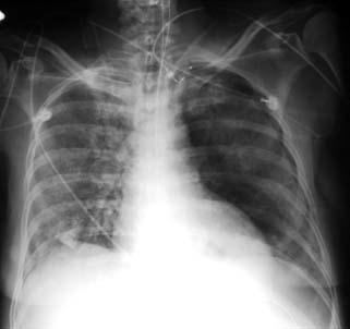
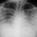
The film now reveals persistent left lower lobe and left upper lobe pneumonia. Note the obliteration of most of the left cardiac border, secondary to the involvement of the inferior division of the lingular segment of the upper lobe. Of interest however, is the pneumothorax, which has developed on the right. This patient has long-standing interstitial fibrosis with a pneumonia now superimposed. As a result of a prolonged period on the respirator, he developed a pneumothorax, which is a common complication.
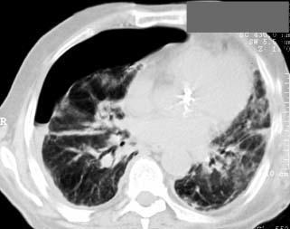
A CT study performed on this patient the following day reveals the extensive right-sided pneumothorax with a small right-sided effusion. The changes in the left lung are seen to better advantage on CT. CT is very sensitive for the evaluation of pneumothorax.
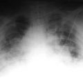
This 42-year-old female was admitted to the ICU, with marked dyspnea and shortness of breath, 2 days after spending a weekend on a farm, working in the barn. Her initial x-rays revealed alveolar densities at the periphery of both lungs, and the diagnosis of a eucenephalic pneumonia was entertained. Her status necessitated that she be intubated. The film was taken 3 days later.
Discussion
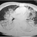
This film reveals the infiltrate in both lung fields in the periphery consistent with an allergic pneumonitis. Note the pneumothorax on the left, secondary to dissection of air out of the alveoli, resulting in a pneumothorax.
Related posts:

Stay updated, free articles. Join our Telegram channel

Full access? Get Clinical Tree


