Base of Thumb Metacarpal Fracture
Ryan E. Embertson
Daniel B. Nissman
CLINICAL HISTORY
39-year-old man presents with wrist pain after a fall from a horse.
FINDINGS
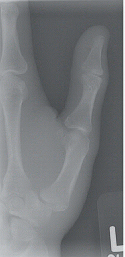
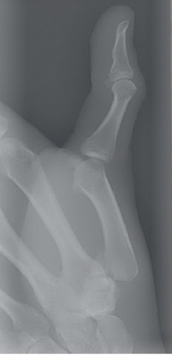
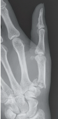
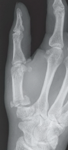
Posteroanterior (PA) (Fig. 64A) and lateral (Fig. 64B) radiographs of the left thumb reveal an oblique intra-articular fracture at the volar medial aspect of the metacarpal base. PA (Fig. 64C) and oblique (Fig. 64D) views of the left hand coned on the thumb in another patient demonstrate an impacted comminuted intra-articular fracture of the thumb metacarpal base; the pattern of comminution can be described as either “T-shaped” or “Y-shaped.”
Stay updated, free articles. Join our Telegram channel

Full access? Get Clinical Tree


