SONOGRAM ABBREVIATIONS
Ao Aorta
Du Duodenum
F Fibroid
GBL Gallbladder
IVC Inferior vena cava
K Kidney
L Liver
P Pancreas
SMA Superior mesenteric artery
Sp Spleen
Spa Splenic artery
Spv Splenic vein
St Stomach
Ut Uterus
KEY WORDS
Acoustic Enhancement. Because sound traveling through a fluid-filled structure is barely attenuated, the structures distal to a cystic lesion appear to have more echoes than neighboring areas. Also referred to as through transmission (information to follow; see and Fig. 5-1).
Anechoic. Without internal echoes. Not necessarily cystic unless there is distal echo enhancement (good through transmission).
Complex. A structure that has both fluid-filled (echo-free) and solid (echogenic) areas.
Contralateral. On the other side of the body.
Cyst. Spherical, fluid-filled structure with well-defined walls that contains few or no internal echoes and exhibits good acoustic enhancement.
Cystic. In ultrasonography, the word cystic does not necessarily refer to a cyst. The term is used (inaccurately) by some to describe any fluid-filled structure (e.g., urine-filled bladder or bile-filled gallbladder; see Fig. 5-1).
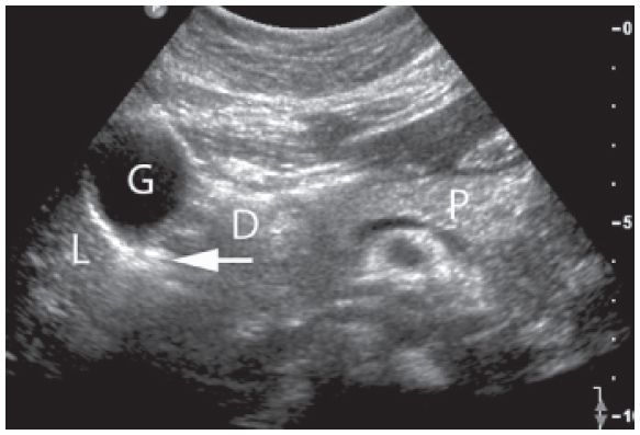
Figure 5-1. ![]() Transverse section of the upper abdomen showing the usual echogenicity of the organs in a young adult. Note that the pancreas (P) contains more echoes than the liver (L). The gallbladder (G), a “cystic” (fluid-filled) structure, shows acoustic enhancement behind it (arrow), in the region of the duodenum (D). The spleen is slightly more echogenic than the liver.
Transverse section of the upper abdomen showing the usual echogenicity of the organs in a young adult. Note that the pancreas (P) contains more echoes than the liver (L). The gallbladder (G), a “cystic” (fluid-filled) structure, shows acoustic enhancement behind it (arrow), in the region of the duodenum (D). The spleen is slightly more echogenic than the liver.
Distal. The extremity (limb) end of a body structure.
Echo-free. See Anechoic.
Echogenic. Describes a structure that produces echoes. Usually a relative term. For example, Figure 5-1 shows the normal texture of the liver and pancreas; the pancreas is slightly more echogenic. A change in the normal echogenicity signifies a pathologic condition.
Echogram. Term used by some to describe an ultrasonic examination, especially in cardiac work; an echocardiogram is frequently referred to as an “echo.”
Echolucent. Without internal echoes; not necessarily cystic.
Echopenic. A few echoes within a structure; less echogenic. The normal kidney is echopenic relative to the liver (see Fig. 5-1).
Echo-poor. See Echopenic.
Echo-rich. See Echogenic.
Fluid-Fluid Level. Interface between two fluids with different acoustic characteristics. This interface has a horizontal level that varies with patient position.
Footprint. Descriptive term for the amount of transducer face in contact with the patient (i.e., a small-head transducer has a small footprint).
Gain. The strength of the echoes throughout the image can be varied by changing the power output from the system.
Homogeneous. Of uniform composition. The normal texture of several parenchymal organs is homogeneous (e.g., liver, thyroid, and pancreas).
Hyperechoic. See Echogenic.
Hypoechoic. See Echopenic.
Interface. Strong echoes that delineate the boundary of organs and that are caused by the difference between the acoustic impedance of two adjacent structures. An interface is usually more pronounced when the transducer is perpendicular to it (Fig. 5-2).
Ipsilateral. On the same side of the body.
Isoechoic. Of the same echogenicity as a neighboring area, but not necessarily of the same texture.
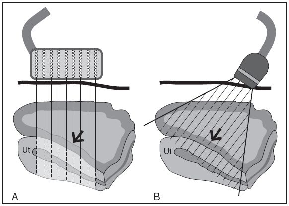
Figure 5-2. ![]() Interface. A. The “interface” between the bladder (black arrow) and the uterus (Ut) is poorly defined because the linear array beam is not perpendicular to the uterine wall. B. The sector beam was angled perpendicular to the interface (black arrow) and is now well seen. The use of a small-headed scanner can bring out interfaces that are oblique to the linear array beam.
Interface. A. The “interface” between the bladder (black arrow) and the uterus (Ut) is poorly defined because the linear array beam is not perpendicular to the uterine wall. B. The sector beam was angled perpendicular to the interface (black arrow) and is now well seen. The use of a small-headed scanner can bring out interfaces that are oblique to the linear array beam.
Noise. Artifactual echoes resulting from too much gain rather than from true anatomic structures.
Proximal. The trunk end of a limb or organ.
Reverberation. An artifact that results from a strong echo returning from a large acoustic interface to the transducer. This echo returns to the tissues again, causing additional echoes parallel to the first.
Ring Down. Extreme form of reverberation artifact that occurs when a long series of echoes caused by a very strong acoustic interface and consequent reverberations are seen.
Scan. Verb: to perform an ultrasound scan. Noun: a sonographic examination.
Shadowing. Failure of the sound beam to pass through an object. This blockage is caused by reflection or absorption of the sound and may be partial or complete. For example, air bubbles in the duodenum allow poor transmission of the sound beam because most of the sound is reflected. A calcified gallstone does not allow any sound to pass through, and shadowing is pronounced (Fig. 5-3). These degrees of acoustic shadowing may help in diagnosis.
Solid (Homogeneous). A mass or organ that contains uniform low-level echoes because the cellular tissues are acoustically very similar.
Sonodense. A structure that transmits sound poorly. A dense structure can attenuate sound so greatly that the back wall is poorly defined (Fig. 5-4). If it is very homogeneous, there may be few or no internal echoes, but the lack of acoustic enhancement and poor back wall help differentiate it from a cystic, echo-free structure.
Sonogenic. Handsome ultrasound image (photogenic), such as a good example of vascular anatomy.
Sonographer. A health professional who has learned how to perform quality sonography and can tailor the examination to individual patients.
Sonologist. A physician who specializes in ultrasonography and has appropriate training.
Sonolucent (Anechoic). Without echoes. Not necessarily cystic unless there is good through transmission.
Specular Reflector. Structure that creates a strong echo because it interfaces at right angles to the sound beam and has significantly different acoustic impedance from a neighboring structure (e.g., diaphragm/liver or posterior bladder wall/bladder).
Static Scan. Not real time. B-scans produced with a fixed-arm system. Obsolete technique.
Texture. The echo pattern within an organ; could be homogeneous or irregular.
Through Transmission. The amount of sound passing through a structure (see Fig. 5-1). Same as acoustic enhancement (mentioned previously).
Transonicity. Term used to indicate the amount of sound passing through a mass or cyst, usually qualified as good or poor. Same as acoustic enhancement.
Trendelenburg. A position in which a recumbent patient is tilted so that the feet are higher than the head.
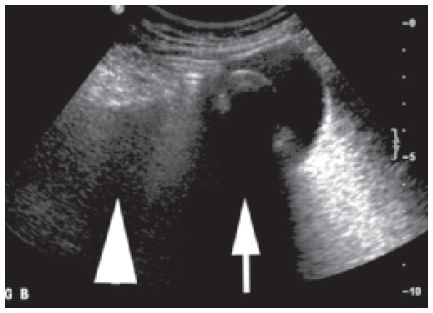
Figure 5-3. ![]() The acoustic shadowing from the stones in the gallbladder (GB) is “sharp” (small arrow), whereas the shadowing from the bowel gas is “soft” (large arrow).
The acoustic shadowing from the stones in the gallbladder (GB) is “sharp” (small arrow), whereas the shadowing from the bowel gas is “soft” (large arrow).
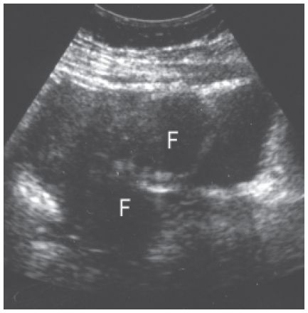
Figure 5-4. ![]() The fibroid (F) at the posterior aspect of the uterus is a solid homogenous mass with some internal echoes. Its density attenuates sound, so the internal echogenicity diminishes near the back wall, and there is poor through transmission.
The fibroid (F) at the posterior aspect of the uterus is a solid homogenous mass with some internal echoes. Its density attenuates sound, so the internal echogenicity diminishes near the back wall, and there is poor through transmission.
Terms Relating to Orientation
ANATOMIC TERMS
See Figures 5-5, 5-6, and 5-7.
Anterior or Ventral. Structure lying toward the front of the patient.
Distal. Away from the origin.
Inferior or Caudal. Terms denoting a structure closer to the patient’s feet.
Lateral. Structure lying away from the midline.
Medial or Mesial. Structure lying toward the midline.
Posterior or Dorsal. Structure lying toward the back of the patient.
Prone. The patient lies on his or her stomach.
Proximal. Nearer to the center of the body.
Quadrant. The abdomen is divided into four quarters, each known as a quadrant.
Superior, Cranial, or Cephalad. Interchangeable terms denoting a structure closer to the patient’s head.
Supine. The patient lies on his or her back.
Terms Relating to Labeling
The American Institute of Ultrasound in Medicine has established standards for labeling studies so that a sonogram done in Columbus, Ohio, can be interpreted with no misunderstanding in Baltimore, Maryland. These standards are occasionally revised and are available on request from The American Institute of Ultrasound in Medicine.
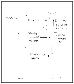
Figure 5-5. ![]() Standard labeling nomenclature used to show where structures lie in relation to each other.
Standard labeling nomenclature used to show where structures lie in relation to each other.
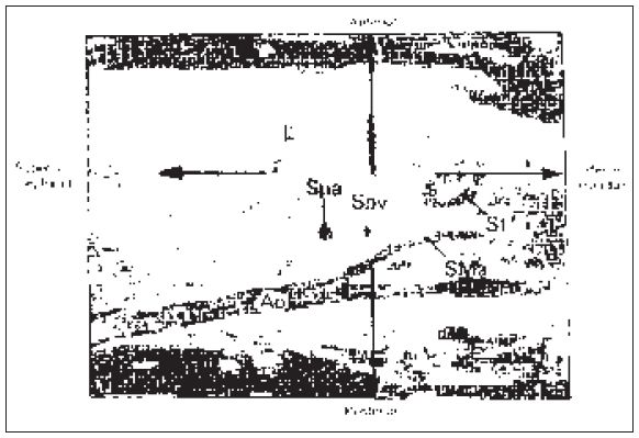
Figure 5-6
Stay updated, free articles. Join our Telegram channel

Full access? Get Clinical Tree


