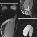
Fig. 2.1
(a, b) Sequential axial T1-weighted fat-suppressed post-contrast images of both breasts. Sagittal T1-weighted fat-suppressed post-contrast image (c), sagittal T1-weighted image (d), sagittal T2-weighted fat-suppressed image (e), and sagittal T1-weighted fat-suppressed image (f) with computer-aided detection (CAD) enhancement kinetics color overlay, all of the right breast
2.2 Intramammary Lymph Node 1
Teaching Points
Intramammary lymph nodes are separate from axillary lymph nodes and are completely surrounded by breast tissue, typically in the upper outer quadrant. Approximately 5 % of patients undergoing mammography and up to 28 % of breast specimens will exhibit these lymph nodes. Characteristic features of a normal intramammary node include a fatty hilum, diameter less than 1 cm, and circumscribed margins. An intramammary lymph node may become abnormal and lose its hilum or enlarge secondary to hyperplasia, metastatic disease, or inflammation. A benign intramammary lymph node demonstrates reniform morphology with a fatty hilum on MRI. The cortex will be T1 hypointense and T2 hyperintense with rapid, homogeneous enhancement; a type III washout kinetic pattern can be seen, as demonstrated in this case.
Image Findings


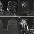
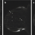
Fig. 2.2
Intramammary lymph node. (a, b) Sequential axial T1-weighted fat-suppressed post-contrast images demonstrate a homogeneously enhancing mass (arrows) in the outer right breast, which appears oval on one slice and reniform on the next. The reniform appearance is characteristic of a lymph node. (c) A sagittal T1-weighted fat-suppressed post-contrast image through the mass highlights its homogeneous enhancement and reniform shape (arrow). The lymph node (arrows) has low signal on the T1-weighted image (d) and high signal on the T2-weighted fat-suppressed image (e). Its central notch (arrowheads) is isointense to fat on both sequences. (f) CAD enhancement kinetics analysis demonstrates a color-coded mixed progressive (yellow) and washout (red) enhancement pattern
2.3 History
34-year-old women high-risk screening MRI (Fig. 2.2).

Fig. 2.3
(a) T1-weighted fat-saturated post-contrast sagittal image of the right breast with CAD kinetic analysis (b). (c) Post-contrast sagittal subtraction image. (d) Corresponding T2-weighted fat-saturated image. (e) T1-weighted sagittal image
2.4 Intramammary Lymph Node 2
Teaching Points
Normal intramammary lymph nodes are usually seen as oval or reniform, smoothly marginated, fat-containing masses. They are often seen adjacent to vessels within the upper outer quadrant, although 28 % of intramammary lymph nodes are found in other locations. They are usually nonpalpable and are found incidentally on breast MRI, where they appear hyperintense on T2-weighted images. They typically demonstrate homogenous enhancement with rapid initial and washout delayed kinetics. Non–fat-saturated T1-weighted images are usually helpful to confirm the presence of fat within the hilum. Biopsy can be avoided if the mass demonstrates classic characteristics of a benign lymph node (Figs. 2.3 and 2.4).
Image Findings

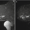
Fig. 2.4
Intramammary lymph node. (a) Sagittal T1-weighted fat-saturated post-contrast image of the right breast demonstrates a 4-mm focus of enhancement (arrow) in the lower outer anterior breast with a suspicious washout enhancement pattern (b) color-coded as red on CAD. (c) On the subtraction image, the morphology of the enhancement appears reniform (arrow) with an adjacent feeding vessel (arrowhead). (d) A sagittal T2-weighted fat-saturated image demonstrates peripheral hyperintensity (arrowhead), and a sagittal T1-weighted image (e) demonstrates central hyperintensity (arrowhead), compatible with a fatty hilum
2.5 History
37-year-old women high-risk screening MRI (Figs. 2.5 and 2.6).


Fig. 2.5
Sagittal T2-weighted fat-saturated image (a) and corresponding sagittal T1-weighted post-contrast subtraction image (b) of the right breast
Fig. 2.6
Sagittal T2-weighted fat-saturated image (a) and corresponding sagittal T1-weighted fat-saturated pre-contrast image (b) of the left breast
2.6 Hemorrhagic and Inflamed Cyst
Teaching Points
Simple cysts result from dilatation and effacement of the terminal duct lobular unit. They are commonly seen incidentally on MRI and follow fluid signal on all sequences (appearing very hyperintense on T2-weighted images and intermediate to hypointense on T1-weighted images). On post-contrast images, cysts typically do not enhance, but the periphery of the cyst may enhance if there is surrounding inflammation. Not infrequently, cysts may have intermediate or high signal on T1-weighted images owing to proteinaceous or hemorrhagic contents (Figs. 2.5, 2.6, 2.7, and 2.8).
Image Findings

Fig. 2.7
Inflamed cyst. Sagittal T2-weighted fat-saturated image (a) and corresponding sagittal T1-weighted post-contrast subtraction image (b) of the right breast demonstrate a T2 hyperintense round mass (arrow) with rim enhancement (arrowhead) in the upper central breast

Fig. 2.8
Hemorrhagic/proteinaceous cyst. Sagittal T2-weighted fat-saturated image (a) and corresponding sagittal T1-weighted fat-saturated pre-contrast image (b) demonstrate a T1 and T2 hyperintense lobular mass in the upper central breast (arrows), compatible with a hemorrhagic cyst. For comparison, note the smaller, more posterior non-hemorrhagic cyst (arrowhead)
2.7 History

2.8 Simple Cyst and Phase-Encoding Artifact
Teaching Points
Phase-encoded motion artifact commonly seen in breast imaging is due to physical movement of the patient and manifests as ghosting in the direction of phase encoding, which is usually in the direction of the short axis. The spacing between the artifacts is related to the TR and frequency of the motion. Unlike Gibbs or truncation artifacts, the motion artifacts usually extend across the entire field of view. Possible solutions to phase-encoded motion artifact include cardiac/respiratory gating, switching phase and frequency encoding directions, increasing the number of signal averages, and shortening the scan time when patient motion is causing the artifact.
Image Findings

Fig. 2.10
Benign cyst and ghosting artifact. Sagittal T2-weighted fat-saturated image of the left breast shows a benign T2 hyperintense 3 cm cyst in the central breast. Ghosting artifact is seen superior and inferior to the cyst as well as superior and inferior to the separate T2 hyperintense nipple marker (arrows)
2.9 History
44-year-old women high risk screening MRI.

Fig. 2.11
Sagittal T1-weighted fat-saturated post-contrast (a) and pre-contrast (b) images. (c) Sagittal post-contrast subtraction
2.10 Dilated Duct
Teaching Points
Normal ducts usually are not well visualized on breast MRI. In the setting of a chronic obstructive process, however, they become dilated and present as high-signal-intensity, branching tubular structures on T1-weighted images and as hypointense structures on T2-weighted images (owing to proteinaceous or hemorrhagic material). Dilated ducts do not typically enhance, but enhancement of the ductal wall may be seen because of underlying inflammation. It is important to remember that assessment of enhancement should not depend solely on the subtracted images, as misregistration artifact may occur (Figs. 2.11 and 2.12).
Image Findings

Fig. 2.12
Benign dilated ducts. (a) Sagittal T1-weighted fat-saturated post-contrast image of the left breast demonstrates linear hyperintensity (arrows) in the central middle third of the breast, which could represent non-mass enhancement. (b) The pre-contrast sequence demonstrates similar linear hyperintensity (arrows). (c) The sagittal post-contrast subtraction image demonstrates no enhancement in this location. Findings are consistent with proteinaceous or hemorrhagic material in the ducts
2.11 History
36-year-old woman with BRCA1 mutation undergoing high-risk screening breast MRI.


Fig. 2.13
(a, b) Sagittal T2-weighted fat-saturated images of the right breast. No suspicious enhancement was present on post-contrast images (not shown)
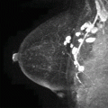
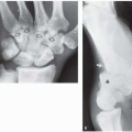
Fig. 2.14
Mediolateral oblique (MLO) (a) and craniocaudal (CC) mammographic (b) images of the right breast
2.12 Dilated Duct with Air
Teaching Points
Image Findings

Fig. 2.15
Dilated duct with air. (a) Magnified sagittal T2-weighted fat-saturated MRI image of the right breast shows linear branching signal voids within subareolar ducts (arrows), representing air. (b) Magnified CC mammographic image of the right breast shows corresponding linear branching lucencies (arrows)
2.13 History

2.14 Dilated Duct with Hemorrhage/Proteinaceous Debris
Teaching Points
Normal ducts usually are not seen on MR imaging, as they blend in with the background parenchyma. In cases of inflammation, infection, or obstruction, however, ducts filled with protein or blood will be visible on non-enhanced T1-weighted images as high-signal-intensity, branching tubular structures without associated enhancement. Therefore, subtraction images are very helpful when pre-contrast images demonstrate a hyperintense ductal pattern, to exclude abnormal enhancement. Otherwise, subtle areas of ductal wall enhancement may be overlooked, and a potential underlying carcinoma can be missed.
Image Findings

Fig. 2.17
Intraductal proteinaceous/hemorrhagic material. (a) Sagittal T1-weighted fat-suppressed pre-contrast image demonstrates segmentally distributed linear high signal (arrows) in the lower breast. Although one could mistake the high signal in this region on the sagittal T1-weighted fat-suppressed post-contrast image (b) for enhancement, the post-contrast subtraction image (c) shows that no enhancement is actually present
2.15 History
30-year-old women high risk screening MRI (Figs. 2.18 and 2.19).


Fig. 2.18
(a) Axial post-contrast subtraction image of both breasts. (b) Magnified image of the left breast from a. (c) CAD overlay. (d) Axial T2-weighted fat-saturated image of both breasts. (e) Magnified image of the left breast from d
Fig. 2.19
Targeted second-look ultrasound image of the left breast
2.16 Fibroadenoma
Teaching Points
Fibroadenomas are frequently seen on breast MRI and are best identified using morphologic features, classically a smooth margin with hypointense internal septa on T2-weighted images. Although less than 40 % of fibroadenomas demonstrate nonenhancing internal septations, if present they can be predictive of benignity (>95 %). MRI enhancement kinetics alone is neither specific nor sensitive because of the overlap in enhancement characteristics between benign and malignant lesions (Figs. 2.18, 2.19, 2.20, and 2.21).
Image Findings

Fig. 2.20
Fibroadenoma on MRI. (a) Axial post-contrast subtraction image shows a homogeneously enhancing oval mass (arrow) along the 8:00 axis of the left breast. (b) The nonenhancing internal septations (arrows) are seen best on the close-up image. (c) CAD overlay demonstrates a progressive pattern of enhancement as evidenced by blue color assignment (arrow). Axial T2-weighted fat-saturated full-field (d) and close-up (e) images demonstrate a corresponding T2 hyperintense oval mass (arrow) with T2 hypointense internal septae (arrows)

Fig. 2.21
Fibroadenoma on ultrasound. Second-look ultrasound was performed, demonstrating an oval, circumscribed, hypoechoic mass corresponding to the MRI mass
2.17 History
17-year-old girl with bilateral breast masses (Figs. 2.22, 2.23, 2.24, and 2.25).


Fig. 2.22
(a) Axial T1-weighted image. (b) Corresponding axial T2-weighted fat-saturated image. (c) Corresponding axial post-contrast subtraction image
Fig. 2.23
More superior axial T1-weighted fat-saturated post-contrast image with CAD kinetics analysis color overlay
2.18 Juvenile Fibroadenomas
Teaching Points
The patient underwent surgical excision of these masses, yielding juvenile fibroadenomas. MRI features in this case are typical for a fibroepithelial lesion, showing circumscribed masses with T1 hypointensity and T2 hyperintensity with nonenhancing septations. Juvenile or cellular fibroadenoma is an uncommon histologic variant of fibroadenoma that frequently undergoes markedly rapid growth. A fibroadenoma over 5–10 cm in diameter is termed a giant fibroadenoma. Approximately 10–25 % of patients with juvenile fibroadenomas have multiple or bilateral tumors at presentation.
Image Findings

Fig. 2.24
Juvenile fibroadenoma. (a) Axial T1-weighted image shows a large, 9.5-cm circumscribed oval hypointense mass occupying most of the left breast. There are areas of linear lower signal intensity (arrows) within the mass, likely representing septations. A smaller 2.2-cm right breast mass with similar signal characteristics is noted. (b) These masses show corresponding T2 hyperintensity with areas of linear higher signal intensity (arrows) within the masses likely representing septations. (c) Axial post-contrast subtraction image shows intense homogeneous enhancement of both masses with central linear areas of non-enhancement corresponding to the T1 hypointense and T2 hyperintense septations

Fig. 2.25
More superior axial T1-weighted fat-saturated post-contrast image with CAD kinetics analysis shows a third mass (4.3 cm, arrow) in the right breast. Both masses demonstrate a predominantly persistent delayed enhancement pattern color-coded as blue
2.19 History
36-year-old with right breast palpable concern (Figs. 2.26, 2.27 and 2.28).


Fig. 2.26
36 Years old women high risk screening MRI. Mammographic images of the right breast. CC (a) and MLO (b) views with corresponding spot compression views (c, d) in a region of palpable abnormality
Fig. 2.27
A targeted right breast ultrasound
Fig. 2.28
Sagittal MRI images of the right breast. T2-weighted fat-saturated image (a) and T1-weighted fat-saturated pre-contrast image (b) and post-contrast image (c). CAD enhancement kinetics overlay (d)
2.20 Phyllodes Tumor
Teaching Points
Phyllodes tumors are rare and constitute 0.3–1.0 % of all breast tumors. They have a high incidence of local recurrence after surgery and potential for hematogenous metastases. Because of their rapid growth, phyllodes tumors tend to have circumscribed margins on mammography and sonography, so it is difficult to distinguish them from fibroadenomas on the basis of imaging. Large phyllodes tumors have a characteristic morphologic appearance on breast MRI, including smooth margins, internal cysts, and septations. They typically demonstrate low T1-weighted signal intensity and appear somewhat bright on T2-weighted images. Literature has shown that contrast enhancement pattern can be variable, so differentiation of phyllodes tumors and fibroadenomas on breast MRI remains unreliable (Figs. 2.26, 2.27, 2.28, 2.29, 2.30, and 2.31).
Image Findings

Fig. 2.29
Phyllodes tumor on mammography. Right CC (a) and MLO (b) views with corresponding spot compression view (c) of the region of palpable abnormality (marked by a BB) demonstrate extremely dense breast tissue without underlying abnormality

Fig. 2.30
Phyllodes tumor on ultrasound. Image from a targeted right breast ultrasound demonstrates a circumscribed, round, homogeneous, hypoechoic mass (between calipers)

Fig. 2.31
Phyllodes tumor on sagittal MRI images. T2-weighted fat-saturated image (a) and T1-weighted fat-saturated pre-contrast image (b) and post-contrast image (c) demonstrate a round, homogeneously enhancing T1 isointense and T2 hyperintense mass (arrows). (d) CAD demonstrates mixed enhancement kinetics (arrow) with a predominant washout pattern
2.21 History
36-year-old woman with discordant biopsy result yielding mastitis (Figs. 2.32 and 2.33).

Fig. 2.32
(a) Selected sagittal T1-weighted post-contrast fat-saturated image of the left breast. (b) Corresponding CAD kinetics image. (c) Selected axial T1-weighted post-contrast fat-saturated image of the left breast. (d) Selected axial T1-weighted post-contrast fat-saturated image of the left axilla
2.22 Breast Abscess 1
Teaching Points
Breast infection/abscess can result from unresolved local infection, obstruction of the duct near the nipple and acute, chronic or lactational mastitis. They are usually palpable, often associated with erythema, edema, and induration or overlying skin and pain. Depending on the water content of the collection, MR T2-weighted signal may vary. The overlying thickened skin may be intermediate or bright in signal depending on the degree of edema. Post-contrast T1-weighted images usually demonstrate a non-enhancing central, round, or irregular mass surrounded by an early, intensely enhancing rim. Abscess may be confused with malignancy due to the enhancement kinetics of the rim and overlying skin thickening, as illustrated in this case. In addition, focal skin thickening and axillary lymphadopathy can also be present. In case of persistent clinical concern with no resolution with antibiotics, surgical management may be appropriate.
Image Findings

Fig. 2.33
Mastitis/abscess. (a) Sagittal image of the left breast shows irregular masses (spiculated mass; arrow) and rim-enhancing mass (arrowheads) in the upper breast. (b) Corresponding suspicious washout kinetics are present in the CAD images. (c) Focal nodular skin thickening is present in the outer anterior left breast (arrow) In the axilla, enlarged lymph node is present (arrow in d). Initial biopsy of the findings in the upper breast yielded inflammatory changes without evidence of malignancy. Following the MRI, surgical excision was performed yielding inflammatory changes without evidence of carcinoma
2.23 History

2.24 Breast Abscess 2
Teaching Points
Breast infection/abscess can result from unresolved local infection, iatrogenic in etiology, obstruction of the duct near the nipple, and acute, chronic or lactational mastitis. Depending on the water content of the collection, MR T2-weighted signal may vary but often is hyperintense. The overlying thickened skin may be intermediate or bright in signal depending on the degree of edema. Post-contrast T1-weighted images usually demonstrate a non-enhancing central, round, or irregular mass surrounded by an early, intensely enhancing rim. All these features are illustrated in this case.
Image Findings

Fig. 2.35
Breast mastitis/abscess. (a) There is an irregular T2-weighted hyperintense collection (black arrowheads) with a thick heterogeneous signal rim (white arrowheads) and thickening and edema of the overlying skin (white arrowhead). Multiple punctate signal voids (black arrows) are present, reflecting gas bubbles. (b) Sagittal T1-weighted fat-suppressed post-contrast image demonstrates thick rim enhancement (white arrows)
2.25 History
36-year-old woman with a strong family history of breast cancer undergoing high-risk screening breast MRI (Figs. 2.36, 2.37, 2.38, and 2.39)