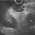
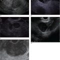
M. Babitha Reddy1, David H. Robbins1, and Mohamad A. Eloubeidi2 1 Lenox Hill Hospital, New York, NY, USA 2 American University of Beirut School of Medicine, Beirut, Lebanon Benign mediastinal lymphadenopathy is a common incidental finding during endoscopic ultrasound (EUS) (Figures 12.1–12.5). Benign enlarged mediastinal lymph nodes can be reactive lymph nodes from exposure to a pulmonary infection, or granulomatous lymph nodes from sarcoid, histoplasmosis, or tuberculosis. Echogenic features include a crescent or triangular shape. Sometimes there is a central hyperechoic strand suggestive of a blood vessel. The borders are irregular and the short axis is often less than 1 cm. There are often multiple lymph nodes. It is not uncommon to encounter the benign “draping” subcarinal lymph node, which can be ovoid, intermediate in echogenicity, and up to 2 cm in length.
12
Benign Mediastinal Lesions
Related posts:


Stay updated, free articles. Join our Telegram channel

Full access? Get Clinical Tree


