Normal intestinal wall thickness depends on the degree of bowel distention and the imaging modality. The normal jejunum wall thickness measures approximately 2 mm and the ileum 1 mm on enteroclysis. On computed tomography (CT), 3 mm is accepted as the upper limit of normal when the bowel is completely distended.
The hallmark of benign wall thickening is homogenous or stratified wall thickening. The appearance is due to low attenuation of the submucosa from edema, inflammation, or fat deposition and is also referred to as a “target” sign. In this chapter, benign causes of small bowel wall thickening ( Box 27-1 ) and benign small bowel neoplasms ( Table 27-1 ) are discussed.
- •
Inflammatory bowel disease (Crohn’s disease, ulcerative colitis [backwash ileitis])
- •
Infectious causes
- •
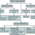
Vascular causes
- •
Miscellaneous causes (eosinophilic enteritis, Whipple’s disease, amyloidosis, graft-versus-host disease, intestinal lymphangiectasia)
- •
Benign neoplasms of small bowel
| Lesion | Age | Sex | Distinguishing Clinical History | Distinguishing Clinical Presentation | Imaging Modality of Choice | Distinguishing Imaging Findings | Enhancement Pattern | Additional Findings |
|---|---|---|---|---|---|---|---|---|
| Benign neoplasms or neoplasm–like lesions | 5th-6th decade | M = F | Could be incidental on imaging | Associated as a spectrum of polyposis syndromes or, if large, can manifest as small bowel obstruction, intussusception, palpable mass. Rarely perforation is noted. | Enteroclysis, wireless capsule endoscopy, CT enterography | Lipomas are fat-containing lesions. GISTs are exophytic masses with hemorrhage, necrosis, and calcification. | Variable | If associated with syndromes |
| Inflammatory bowel disease | 15-25 yr | M = F, four times more common in Ashkenazi Jews | Right lower quadrant pain, intermittent diarrhea, weight loss, rectal bleeding | Perianal fistula formation, extraintestinal manifestations | Enteroclysis and wireless capsule endoscopy for subtle mucosal involvement CT or MR enterography for extent of the disease and complications | Asymmetric and discontinuous involvement Coarsening of villous pattern and wall stratification Linear/aphthous ulcers Cobblestoning String sign secondary to inflammation and spasm Sinus tracts/fistulas and abscess formation | Fibrofatty proliferation, variable enhancement | Extraintestinal manifestations such as sclerosing cholangitis, joint involvement, etc. |
| Infectious diseases | Any age, more common in pediatric patients | M = F, endemic in some places | Immune suppression, recent history of travel or living in endemic places | Diarrhea, acute or chronic manifestation, fever, abdominal pain | Usually none; in chronic cases or immunocompromised patients CT may be helpful. | Nonspecific findings; usually terminal ileum and right colon are involved. | Variable, diffuse enhancement of the involved bowel segment | |
| Vascular diseases | More common in elderly patients | M = F | History of atherosclerotic disease, blood dyscrasia, abdominal surgery or infection | Abdominal pain, acute abdomen, gastrointestinal bleeding | CT; CT or MR angiography | Thrombus in the mesenteric vessels Intestinal wall thickening, “thumbprinting” Pneumatosis | Decreased or sometimes increased enhancement of the bowel wall | Atherosclerotic disease |
Crohn’s Disease
Etiology
Crohn’s disease is an idiopathic, transmural inflammatory process that can affect the entire gastrointestinal tract, with a tendency toward segmental distribution. Environmental factors, microbial influences along with immunologic dysregulation, and genetic factors have been implicated as the cause of this disease.
Prevalence and Epidemiology
Crohn’s disease is more common in northern Europe and North America, and its incidence has increased in recent years and then reached a plateau. The disease most commonly affects young adults between 15 and 25 years of age. A second peak in the elderly is thought to be caused by unrecognized ischemic colitis. Crohn’s is two to four times more common in the Jewish population. The prevalence of disease is increased in the relatives of those who have the disease. For patients who have Crohn’s disease, the occurrence of disease in their offspring is 9.2%. However, classic mendelian inheritance patterns are not seen and Crohn’s disease cannot be linked to a single gene locus.
Clinical Presentation
The typical manifestation is recurrent episodes of abdominal pain, diarrhea, and low-grade fever. The pain is usually in the right lower quadrant and steady. Cramping abdominal pain can suggest intestinal obstruction. If there is colonic involvement, rectal bleeding and perianal fistulas may develop. During the advanced stages of the disease, stricture formation and partial small bowel obstruction are common. Free perforation of the small bowel into the peritoneal cavity is rare, but small, sealed-off perforations are characteristic of the disease and may lead to fistula formation. The risk for colorectal cancer is 4 to 20 times higher than that of the general population.
Extraintestinal manifestations are more common in Crohn’s disease than in ulcerative colitis. Biliary complications are the most common and include cholelithiasis and sclerosing cholangitis. Urolithiasis, sacroiliitis, peripheral arthritis, and ocular and skin manifestations are the other systemic complications, which can be seen in 20% to 30% of the patients.
Anatomy
Crohn’s disease can involve any part of the gastrointestinal tract in a discontinuous fashion, with the terminal ileum and colon most frequently affected. Isolated small bowel involvement can occur in one third of patients. The jejunum and ileum (sparing the terminal ileum) are affected by the disease in 3% to 10% of the patients.
Pathology
The classic gross description of Crohn’s disease is segmental, transmural inflammation of the bowel wall with skip lesions. The mucosa shows multiple aphthous or linear ulcers, and the intervening normal mucosa has the appearance of pseudopolyps. Noncaseating granulomas are not pathognomonic but are very suggestive of the disease; they are found only in up to two thirds of biopsy specimens. Fissures and fistula tracts are common, and the serosal surface may show transmural inflammation associated with a thickened layer of surrounding fat that is also referred to as fibrofatty proliferation.
Imaging
Radiography
Plain radiographs of the abdomen are obtained in patients with acute presentation to evaluate for intestinal obstruction or perforation. Incidental findings may include gallstones and urinary stones. In cases of severe disease, wall thickening of the bowel segments also may be apparent on plain radiographs.
In a prospective trial, Lee and colleagues showed that CT and magnetic resonance (MR) enterography and small bowel follow-through (SBFT) are equally accurate in detecting inflammation in the small intestine. The sensitivity values of CT enterography (89%) and MR enterography (83%) were slightly higher than those of SBFT (67% to 72%). CT and MR were more accurate in depicting extraintestinal complications such as sinus tracts, fistulas, and abscesses. The addition of peroral pneumocolon to the SBFT allows better evaluation of the terminal ileum. A wireless video capsule endoscopy is superior in the detection of early nonstricturing small bowel Crohn’s disease but is contraindicated in stricturing disease.
Coarsening of the villous pattern and thickening of the intestinal folds secondary to edema and inflammation can be seen in early disease. Ulcerations can be aphthous or linear. Aphthous ulcers are punctuate, shallow, discrete depressions surrounded by a halo. Linear ulcers can be long and run parallel to the mesenteric border ( Figure 27-1 ). Together with mesenteric border shortening and antimesenteric sacculations, these ulcers are very characteristic for Crohn’s disease ( Figure 27-2 ). Linear ulcers may penetrate the entire wall of the bowel. Islands of intervening normal mucosa surrounded by denuded mucosa can give the appearance of pseudopolyps, and multiple polypoid elevations can produce the “cobblestoning” appearance ( Figure 27-3 ).


Stenosis of the small intestine in Crohn’s disease can be a combination of fibrosis, inflammation, and spasm. The “string” sign represents intense spasm of the bowel and indicates transmural inflammation ( Figure 27-4 ). This needs to be differentiated from strictures secondary to fibrosis that are not distensible and cause obstruction ( Figure 27-5 ). Asymmetric and discontinuous involvement of the bowel wall are the other hallmarks of Crohn’s disease. Complications such as sinus tracts, fistulas, and abscesses either ending blindly or penetrating the colon or another organ can be visualized by SBFT or enteroclysis ( Figures 27-6 and 27-7 ).


Computed Tomography
CT can be used to diagnose, monitor therapy, and evaluate complications related to Crohn’s disease. When performed with neutral oral contrast agents, it allows mucosal evaluation. CT features of active small bowel Crohn’s disease are bowel wall thickening, mural hyperenhancement, and mural stratification ( Figure 27-8 ). Narrowing of the lumen is another common finding, which can be fixed (as a result of fibrosis) or reversible (inflammation or spasm) ( Figure 27-9 ). Enlarged vasa recta and increased amount of fat surrounding the involved bowel segments are common findings ( Figure 27-10 ). Fistulas between the small bowel and colon or other organs can be seen as enhancing tracts ( Figure 27-11 ). Multiplanar reformatted views may better assist in depiction of fistulous tracts. Positive oral contrast agents may be preferred if a fistula is clinically suspected. In a prospective study by Hara and colleagues, Crohn’s disease was depicted by CT enterography in 53%, ileoscopy in 65%, SBFT in 24%, and capsule endoscopy in 71% of the patients.


Magnetic Resonance Imaging
MR enterography can provide a systematic evaluation of the entire small bowel and the mesentery without exposing the patients to ionizing radiation. In a study by Schreyer and colleagues, all pathologic findings seen on conventional enteroclysis were seen on MR enterography or MR enteroclysis. Some reports have shown greater than 90% sensitivity and specificity with MRI in detection of Crohn’s disease. Increased enhancement and thickness of the bowel wall suggest active inflammation ( Figure 27-12 ). The inflamed segments have increased perfusion and show restricted diffusion.

High signal intensity in the bowel wall on T2-weighted images also indicates active disease, and low signal on T2-weighted images with homogenous, cordlike enhancement is suggestive of chronic Crohn’s disease.
Ultrasonography
During the past decade, advances in the resolution of ultrasound technology with development of high-frequency probes, harmonic imaging, and improved temporal resolution have led to better imaging of the bowel wall. Common ultrasonographic findings of Crohn’s disease are thickened bowel wall and decreased peristalsis. Wall thickness in the range of 3.5 to 15 mm is considered pathologic, with amount of thickening influencing the sensitivity and specificity of detecting inflammation, ranging from 75% to 94% and 67% to 100%, respectively.
Despite the high sensitivity of ultrasonography in the diagnosis of Crohn’s disease, ultrasonography underestimates the extent of involvement. Use of oral and intravenous contrast agents may further increase the diagnostic capability of ultrasonography.
Nuclear Medicine
Labeled white blood cell scans (indium-111 tropolone or technetium-99m hexamethyl propylene amine oxime [ 99m Tc-HMPAO]) and granulocyte scintigraphy with labeled antibodies have been reported to detect the inflammation in Crohn’s disease. Reported sensitivities vary between 5% and 70%, and they have been suggested to be more useful in reassessments rather than the initial diagnosis.
Positive Emission Tomography With Computed Tomography
Recently, fluorodeoxyglucose (FDG)-labeled positron emission tomography (PET) combined with CT (PET/CT) has been shown to be a reliable tool for detection of active disease in both the small and large bowel ( Figure 27-13 ). In a study comparing PET, MR enterography, and granulocyte scintigraphy with labeled antibodies, FDG-PET showed significantly higher sensitivity (85.4%) compared with the other imaging tools for the detection of inflamed bowel segments.

Imaging Algorithm
In patients with suspected Crohn’s disease, enteroclysis, SBFT, and CT or MR enterography can be the initial test, depending on the experience of the radiologist. Capsule endoscopy can be used in patients without stricturing small bowel Crohn’s disease. Radiologic tests also can be helpful to exclude stenosis before the capsule endoscopy and provide more accurate localization of the pathologic process after the capsule endoscopy. Currently, CT enterography is the initial test for patients with acute presentation and suspected obstruction, fistula, or abscess ( Table 27-2 ; see also Figure 27-28 ).
- •
Cobblestoning: Radiographic appearance caused by islands of normal mucosa (appearing as pseudopolyps) surrounded by denuded affected mucosa
- •
“String” sign: Stenosis of the bowel caused by inflammation and spasm
- •
Fibrofatty proliferation: Increased amount of fat around the involved bowel segments
- •
Wall stratification: Low density between mucosal and serosal enhancement representing fat or inflammation
| Modality | Limitations | Pitfalls |
|---|---|---|
| Radiography | Extraintestinal findings, detection of disease activity, radiation | |
| CT | Radiation, insensitive to early mucosal findings | Undistended bowel |
| MRI | Expensive, duration of the test, motion artifacts | Undistended bowel |
| Ultrasonography | Unable to image the entire bowel | Nonspecific findings |
| Nuclear medicine | Radiation, poor spatial resolution | Normal bowel uptake |
| PET/CT | Expensive, radiation, relatively less data available | Normal bowel uptake |
Differential Diagnosis
The early symptoms of Crohn’s disease are mild and nonspecific. Infectious enteritis can radiologically mimic Crohn’s disease. Irritable bowel syndrome and lactose intolerance can have similar clinical presentation. In patients without small bowel involvement, differentiation from ulcerative colitis may be difficult. In elderly patients, ischemic enteritis should be differentiated from Crohn’s disease. Other differential diagnoses include ischemia, neoplasm (lymphoma and, rarely, adenocarcinoma), ulcerative colitis, radiation therapy, vasculitis, and, in children, lymphoid hyperplasia.
Treatment
Medical Treatment
Medical therapy is usually individualized and requires a combination of different drugs to achieve sustained response. The choice of drugs varies according to the severity of the disease, which is based on clinical, biochemical, endoscopic, and histologic findings. The Crohn’s Disease Activity Index (CDAI) score is commonly used to determine the severity of the disease. Different drugs used for Crohn’s disease range from sulfasalazine, to antibiotics in mild cases, to oral corticosteroids, azathioprine, methotrexate, and anti–tumor necrosis factor (TNF) antibody in moderate and severe cases. The use of anti-TNF antibody has revolutionized the therapy of severe Crohn’s disease.
Surgical Treatment
Failure to respond to medical management, development of complications (obstruction, abscess, fistula, or stricture), and inability to tolerate effective therapy are the most common indications for surgical therapy.
- •
Differentiation of Crohn’s disease from other causes of enteritis
- •
Differentiation between active and chronic Crohn’s disease
- •
Recognition of complications
- •
Monitoring of therapy
Infectious Causes
Infectious diseases of the small bowel can be caused by various organisms, including bacteria, viruses, parasites, and fungi. Radiologic signs are often nonspecific, and bowel wall thickening is a common finding. Clinical information such as stool culture, the immune status, and geographic location is helpful for a specific diagnosis.
Etiology
Common bacterial causes of community-acquired infectious enterocolitis in the United States are Campylobacter jejuni, Salmonella, Shigella, Escherichia coli, and Yersinia. Rotavirus and Norwalk virus are the most common pathogens in the children. Adenovirus and cytomegalovirus (CMV) should be considered in immunocompromised patients. Giardia lamblia is the most frequent cause of parasitic enteritis in the United States. Other common parasitic infections that can affect the small intestine are Ancylostoma, Ascaris, Cryptosporidium, and Taenia. Cryptosporidium is a particularly common pathogen in the immunocompromised hosts. With the increasing population of immunocompromised patients (e.g., those with acquired immunodeficiency syndrome), the incidence of Mycobacterium tuberculosis and Mycobacterium avium complex (MAC) has increased during the past 2 decades.
Prevalence and Epidemiology
Acute diarrhea is one of the most common diagnoses in general practice. Immune status, clinical setting, and geographic location are important factors in disease expression and treatment. Acute enteritis causes 3 to 6 million deaths (mostly of children) worldwide. Each year in the United States tens of millions of people acquire gastrointestinal infections with thousands of hospitalizations and deaths. Chronic enteritis occurs less frequently and can be seen with parasites and, less commonly, with bacteria. Immunosuppressed individuals cannot clear pathogens effectively and can develop chronic diarrhea. Campylobacter and Salmonella can cause persistent diarrhea in patients with human immunodeficiency virus infection.
Acute infectious diarrhea is acquired mostly through the fecal-oral route or ingestion of contaminated food and water. In developing countries, infectious enteritis can be endemic; and in most parts of the world, seasonality is recognized in the incidence of acute diarrhea.
Clinical Presentation
Patients may present with abdominal pain, diarrhea (with or without blood), and fever. If the host is a child, elderly, or immunocompromised, dehydration may be frequently encountered. Enteropathogens may involve the entire small bowel, although certain pathogens are more likely to colonize at certain segments. For example, M. tuberculosis tends to involve the terminal ileum and ileocecal valve and Giardia primarily causes duodenal and proximal jejunal disease.
Pathology
In most cases, a pathogen enters and colonizes in an area of intestine. However, ingestion of the toxin alone can cause infection (Staphylococcus aureus, Clostridium botulinum) . Most bacteria disrupt mucosal integrity through cytotoxic mediators. Shigella and enteroinvasive E. coli can cause significant tissue invasion and destruction of the bowel mucosa. Rotavirus may disrupt the mucosa and produce villous atrophy. CMV produces characteristic nuclear and cytoplasmic inclusions that can be recognized on light microscopic examination. Whereas most infections elicit an inflammatory response, parasites such as Giardia or Cryptosporidium cause minimal mucosal response, and it may be difficult to localize these organisms in the villi.
MAC includes two related organisms. M. avium and Mycoplasma intracellulare enter into the intestinal mucosa and are phagocytosed by histiocytes, which are unable to digest them. Acid-fast bacilli within the histiocytes or in the stool samples can be identified. The pathologic findings resemble those of Whipple’s disease, but the intestinal biopsy samples contain non–acid-fast granules and histiocytes are typically foamy. Pathologic findings of infection with M. tuberculosis is similar to that of MAC, with the addition of caseation granulomas in the wall or in the mesentery of the bowel.
Endoscopic findings in infectious enteritis range from normal intestine (mostly viral infections) to inflammation, atrophic or blunted villi, erosions, and ulcers.
Imaging
Radiography
Plain radiographic findings are often normal or nonspecific, showing mild ileus. Barium studies are rarely indicated for acute disease. If the course is prolonged, it is important to differentiate an infectious cause from inflammatory, neoplastic, and vascular causes. The diagnosis depends on the findings of biopsy, stool examination, and culture.
The terminal ileum is most severely affected in infections with Campylobacter and Yersinia, and demonstrates wall thickening with nodular folds and sometimes aphthous ulcers. In Yersinia infection, the bowel mostly retains its normal caliber. These changes also can extend to the cecum and ascending colon. In salmonellosis, barium studies are rarely indicated, and findings are nonspecific with aphthous ulcers and wall thickening most commonly in the region of the terminal ileum. Shigellosis, on the other hand, predominantly colonizes the colon and affects the small intestine by its enterotoxin.
M. tuberculosis causes transaxial ulcerations, polyps, and thickening of the folds mostly in the ileocecal region in the early phase. Involvement is more prominent in the cecum compared with the terminal ileum, and strictures may develop during the course of the disease. Strictures are usually short and have an hourglass configuration and sometimes cause small bowel obstruction. The cecum and ileocecal valve may be unrecognizable, with cephalad retraction of the cecum and straightening of the ileocecal angle. Differentiation of ileocecal tuberculosis from Crohn’s disease is difficult.
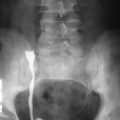
In the immunocompromised host, MAC, Cryptosporidium , and CMV are the most common infective agents causing enteritis. On barium studies, a diffuse granular pattern secondary to nodular thickening of mucosal folds is a common finding in MAC infection. Ulcers are typical findings of CMV enterocolitis and can be large. The ileocecal region is the most commonly involved area, and changes can extend into the cecum and the rest of the colon ( Figure 27-14 ). The barium study findings of cryptosporidiosis are nonspecific fold thickening and increase in intraluminal fluid.
Computed Tomography
CT is usually not indicated in the evaluation of immunocompetent patients with acute enteritis, but it is important to know the CT findings to be able to consider acute enteritis in the differential diagnosis when these findings are incidentally encountered. Nonspecific wall thickening, mild ileus secondary to altered mobility of the involved small bowel segments, and mesenteric adenopathy are the common, nonspecific CT findings of acute enteritis.
CT can demonstrate the extent of the disease in patients with M. tuberculosis infection. CT findings include significant wall thickening mostly involving the ileocecal area, mesenteric adenopathy, and inflammatory masslike lesions in the right lower quadrant. CT also can show ascites and peritoneal and omental soft tissue densities representing peritonitis and may mimic peritoneal carcinomatosis ( Figure 27-15 ).
In the immunocompromised host, CT can help in differentiation of typhlitis (which requires medical treatment) from acute appendicitis and save the patient from unnecessary surgery. CT features of typhlitis include segmental bowel wall thickening involving the terminal ileum, appendix, cecum and ascending colon, pneumatosis coli, and pericolonic fat stranding ( Figure 27-16 ). The extent of the colonic involvement is more substantial in typhlitis, and the presence of known risk factors favors the diagnosis of typhlitis (neutropenic colitis). In patients with MAC infection, detection of mesenteric adenopathy with low attenuation centers indicating necrosis is very suggestive of the cause ( Figure 27-17 ). Similar lymph nodes can be seen in patients with Whipple’s disease in the immunocompetent host.
Magnetic Resonance Imaging
Limited experience is available on the MRI findings of infectious enteritis, and the role of MRI in the diagnosis of small bowel infections has not yet been established.
Ultrasonography
Acute infectious ileitis may show thickening of the ileal wall and mesenteric adenopathy. An involved segment is usually aperistaltic, and the inflammation also can involve the cecum. Demonstration of the normal appendix on ultrasonography can rule out appendicitis.
Nuclear Medicine and Positron Emission Tomography With Computed Tomography
The roles of nuclear medicine and PET/CT have not yet been established in the diagnosis of small bowel diseases.
Imaging Algorithm
Imaging usually is not indicated in immunocompetent patients with acute enteritis (see Figure 27-28 ). Clinical and laboratory evaluation with stool examination with or without cultures is usually enough to make the diagnosis or at least decide about the management. CT may be helpful to rule out a surgical diagnosis or complication of an infectious process. CT is also commonly used for the evaluation of immunocompromised patients with suspected enteritis to identify the cause and the extent of the disease as well as to exclude complications or neoplastic cause ( Table 27-3 ). SBFT and enteroclysis can be obtained in chronic cases. Specific radiologic findings, location, and extent of the disease can help in the accurate diagnosis when evaluated together with the clinical and laboratory information.
| Modality | Accuracy | Limitations |
|---|---|---|
| Radiography | Nonspecific findings and cannot evaluate the extramural disease | |
| Computed tomography | Very helpful in evaluation of acutely presenting immunocompromised patients | Nonspecific findings and cannot evaluate the mucosal disease |
Differential Diagnosis
The clinical diagnosis of infectious enteritis is usually not difficult, although the specific pathogen cannot be determined in all cases. When the inflammation causes ileus, clinical presentation may mimic bowel obstruction. When the course of the infection is chronic, inflammatory, neoplastic, and vascular causes also should be considered in the differential diagnosis.
When there is involvement of the ileocecal area, the differential diagnosis includes Crohn’s disease, radiation enteritis, neoplasm (lymphoma, adenocarcinoma, carcinoid), or extrinsic inflammatory masses such as abscess/phlegmon from acute appendicitis. When the folds are thickened without narrowing and the history is more acute, infection with Yersinia, Salmonella, or Campylobacter is the most likely cause. Luminal narrowing and mesenteric border ulcers are suggestive of Crohn’s disease. Tuberculosis can mimic Crohn’s disease with skip lesions but affects the right colon more than the terminal ileum. In tuberculosis, the ileocecal valve is patulous. Behçet’s disease also has predilection for the terminal ileum, and the presence of other clinical manifestations helps make the diagnosis.
When the involvement is more proximal (jejunum and proximal ileum), ulcerative jejunoileitis, eosinophilic enteritis, lymphoma, and abetalipoproteinemia can be considered in the differential diagnosis. Ulcerative jejunoileitis is a rare complication of celiac disease and may manifest as ulcer formation, which may eventually lead to stricture formation. Radiologically, it may be impossible to differentiate it from lymphoma. In the immunocompromised host, the main differential considerations are neoplasm (lymphoma, Kaposi’s sarcoma) and graft-versus-host disease (GVHD).
Treatment
Medical Treatment
The treatment of infectious enteritis depends on the causative agent. Treatment is mostly hydration and diet alterations because most cases of community-acquired infectious enteritis are self-limiting in the immunocompetent host. In contrast, microorganisms such as most parasites, M. tuberculosis, and Shigella are treated with the appropriate antibiotics. Agent-specific antimicrobial therapy is more aggressively pursued in the immunocompromised host.
Surgical Treatment
Surgical therapy is rarely indicated. It is performed mostly for the complications of intestinal tuberculosis such as perforation, obstruction, or massive hemorrhage.
Stay updated, free articles. Join our Telegram channel

Full access? Get Clinical Tree


