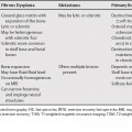
143 Thoracic and lumbar compression fractures are common in patients with senile, steroid-induced or other etiologies of osteoporosis. Metastatic disease also commonly affects the spine and can also result in compression fractures. Some findings on conventional imaging have been shown to be useful discriminating features in differentiating benign from malignant compression fractures (Table 143.1). Signal intensity on T1- or T2-weighted magnetic resonance images (T1WI, T2WI) or enhancement characteristics have not been shown to be useful. Benign and metastatic compression fractures can have diffuse paraspinal masses, but a focal paraspinal mass is more common in metastatic compression fractures. Likewise, an epidural mass that encases the dural sac is more common in metastatic disease.
Benign versus Pathologic (Neoplastic) Fracture
| Malignant | Benign | |
|---|---|---|
| Vertebral Body Posterior Border | Convex posterior border | Retropulsed posterior bone fragment |
| Epidural Mass | May be large (particularly if encasing dural sac) | May be present |
| Paraspinal Mass | May be large (a useful feature, particularly if focal) |