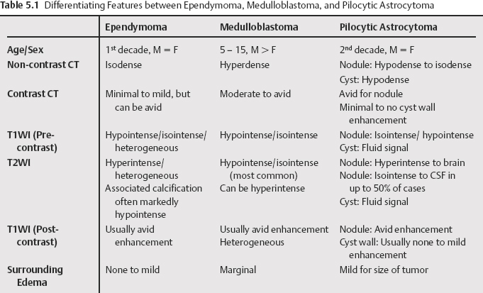
5 The majority of posterior fossa neoplasms in children are intraaxial with pilocytic astrocytomas and medulloblastomas as the most common types. These two tumors combined account for roughly two thirds to three quarters of posterior fossa neoplasms in children. Ependymomas are the third most common posterior fossa tumor, accounting for up to 10%. Medulloblastomas are also categorized as primitive neuroectodermal tumors (PNET). Pilocytic astrocytomas are typically well-delineated, cystic tumors arising in the cerebellar hemispheres or less frequently, the vermis. Medulloblastomas are often well-circumcised, homogeneous, hyperdense vermian masses projecting into the 4th ventricle. Ependymomas are frequently calcified, isodense 4th ventricular masses with a tendency to spread through the foramina of the 4th ventricle. There are features that can aid in the differentiation of these tumors (Table 5.1). In addition to conventional imaging, techniques such as apparent diffusion coefficient (ADC) measurement can serve to differentiate these tumors. A study evaluating the efficacy of ADC in differentiating cerebellar tumors did not find any overlap between the major tumor types (pilocytic astrocytoma, ependymoma, medulloblastoma). Magnetic resonance (MR) spectroscopy has also shown promise in differentiating neoplasms, and when combined with hemodynamic imaging, also may aid in differentiating neoplasm from normal tissue, treated tumor, and areas of necrosis, and may be helpful in evaluating tumor response to therapy as well.
Posterior Fossa Neoplasms in Children
Radiology Key
Fastest Radiology Insight Engine