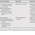
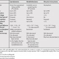
114 The differential diagnosis of ovarian lesions with hyperintensity on T1-weighted magnetic resonance imaging (T1WI) is limited. Teratomas are the only lesions that have T1WI hyperintensity secondary to fat and are easily differentiated with fat-saturated images. Endometriomas are sometimes difficult to differentiate from hemorrhagic cysts. Certain imaging features may be helpful (Table 114.1). The types of T1WI hyperintense ovarian lesions include1
Ovarian Lesions with Hyperintensity on T1-Weighted Magnetic Resonance Imaging