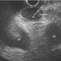
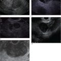
Kapil Gupta Cedars‐Sinai Medical Center, Los Angeles, CA, USA Endoscopic ultrasound provides excellent imaging of the biliary tree and gallbladder. Using radial and linear echoendoscope, visualization of the bile duct is performed from two main stations: from the duodenal bulb and from the second portion of the duodenum. The gallbladder is usually seen from the duodenal bulb or the antrum of the stomach. As the entire biliary tree can be visualized only using linear echoendoscope, more endosonographers are primarily using linear echoendoscope for evaluating bile duct anatomy. The echoendoscope is advanced through the pylorus into the duodenal bulb. To achieve this, the scope is usually in a long position along the greater curvature. The big wheel is then turned downwards and the scope is either turned slightly clockwise or the small wheel rotated slightly to the right to deflect the tip of the echoendoscope towards the apex of the duodenal bulb and impacted there. To achieve a stable position the balloon can be inflated, which helps in maintaining the tip of the scope in the bulb. Here, with slight right and left movement, the liver is oriented at 12 o’clock. In this position the portal vein and the common bile duct can be visualized as two parallel tubular structures to the left of the transducer (Figure 5.1). With slight right rotation the bile duct can be followed towards the head of the pancreas into the ampulla where a thin pancreatic duct can be visualized below the bile duct (Figure 5.2). With left rotation or slight counterclockwise rotation the bile duct can be followed upwards into the liver (Video 5.1). The gallbladder is usually visualized along the under surface of the liver (Figure 5.3) and with slight rotation, withdrawing, and pushing the echoendoscope the entire gallbladder can be visualized (Video 5.1). To visualize the bile duct from the second portion of the duodenum, the transducer is placed along the ampulla and with slight movement the bile duct and the pancreatic duct can be visualized as two round anechoic structures (Figure 5.4) (Video 5.1). The bile duct is visualized with a linear echoendoscope from either the duodenal bulb or the second portion of the duodenum. The transducer of the echoendoscope is advanced across the pylorus. Once in the bulb the balloon can be filled slightly with water to maintain a stable position. Initially the tip of the echoendoscope is impacted at the apex of the bulb; to achieve this, the big wheel is turned downwards. With slightly counterclockwise torque and turning the big wheel down further the bile duct is visualized as a round structure, right in the center of the field (Figure 5.5). The cut section of the visualized bile duct is in the region of the common bile duct or the common hepatic duct. In this view the portal vein is usually visualized below the bile duct, and also the inferior vena cava (IVC) can be seen to the right of the field (Figure 5.5). The common hepatic artery can also be visualized coursing in between the bile duct and the portal vein towards the left of the bile duct. From this view the inward impacted position of the tip of the transducer is maintained. By rotating the transducer in a counterclockwise manner the bile duct is followed towards the hilum into the liver and the bifurcation at the porta hepatis can be visualized. Cystic duct take‐off can also be visualized by tracing the bile duct. Undoing the counterclockwise rotation, and rotating the scope to the right, the transducer is rotated to follow the bile duct in the intrapancreatic portion towards the papilla. With this maneuver the entire bile duct can be followed. In most of the instances the bile duct can be traced downstream all the way to the point where it joins the pancreatic duct into the ampulla. Just below the bile duct, in the intrapancreatic portion, the pancreatic duct can be visualized (Video 5.1). Figure 5.1 Bile duct as visualized from the duodenal bulb (radial echoendoscope). Figure 5.2 Bile duct followed towards the head of the pancreas from the duodenal bulb (radial echoendoscope). CBD, common bile duct; PD, pancreatic duct. Figure 5.3 Gallbladder from the duodenal bulb (radial echoendoscope). CBD, common bile duct. Figure 5.4 Bile duct and pancreatic duct from the second portion of the duodenum (radial echoendoscope). CBD, common bile duct; PD, pancreatic duct. Figure 5.5 Common hepatic duct and cystic duct as visualized from the duodenal bulb (linear echoendoscope). For visualizing the gallbladder the transducer is impacted in the bulb and the big wheel is turned up and the scope rotated counterclockwise so the transducer now faces the undersurface of the liver; by turning the small wheel to the right and left the gallbladder can be seen in the subhepatic region. Moving the big wheel up and down, and turning the small wheel right and left, the entire gallbladder can usually be scanned. Sometimes the scope is slightly withdrawn to visualize the entire gallbladder (Video 5.1). Care should be taken when advancing the echoendoscope into the second portion. By slight inward push, turning the big wheel down and the small wheel to the right, the scope tip usually points in the axis of the second portion of the duodenum. Carefully moving the big wheel up and down the scope tip falls into the second portion of the duodenum. At this point the small wheel is kept turned towards the right and the scope is reduced to a short position to align the transducer along the papilla. Figure 5.6 Bile duct and pancreatic duct from the second portion of the duodenum (linear echoendoscope). The transducer is kept in close apposition with the major papilla. With slight right and left torque while withdrawing the scope, the bile duct is visualized as a long tubular structure closer to the duodenal wall, and deep to it lays the pancreatic duct (Figure 5.6). The bile duct can then be followed towards the liver with continued slow withdrawal of the scope and slight right and left torque. Usually the bile duct can be followed to the common hepatic duct portion when the scope tends to slip back into the stomach; to prevent this scope is again pushed inwards with counterclockwise rotation to gain a position similar to one in the duodenal bulb (Video 5.1). Combining views from both the stations, namely the duodenal bulb and the second portion of the duodenum, the entire biliary tree can be visualized.
5
Bile Duct: Radial and Linear
Normal bile duct anatomy
Normal anatomy of the bile duct and gallbladder with radial echoendoscope
Normal anatomy of the bile duct and gallbladder with linear echoendoscope


Related posts:


Stay updated, free articles. Join our Telegram channel

Full access? Get Clinical Tree


