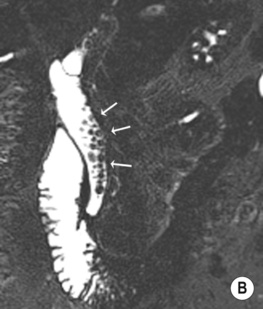
• Heavily T2-weighted coronal oblique fast spin-echo sequence to obtain source data (aligned along the plane of the common bile duct (CBD)) • Source data allows MIP reformats to be generated (highlighting fluid-filled structures) – usually a number of coronal MIP reformats over 180° • Secretin: this stimulates exocrine pancreatic secretion, distending the pancreatic duct and improving its visualization (acts immediately, returning to baseline at 10 min) • Functional MR cholangiography: using delayed imaging at 30–60 mins with the hepatobiliary excreted contrast agents Gd-EOB-DTPA (Primovist) or Gd-BOPTA (MultiHance) • Normal morphology: only central intrahepatic ducts are normally seen (≤ 3mm) • Right posterior hepatic duct (segments VI/VII): almost horizontal course • Right anterior hepatic duct (segments V/VIII): more vertical course • Left hepatic duct (segments II–IV): joins the right to form the common hepatic duct • Cystic duct insertion into common hepatic duct: right lateral (50%) • Common variants: an aberrant right posterior duct draining into the common hepatic duct or cystic duct • Technique: volume averaging artefacts in MIP reformats can obscure filling defects – source images must always be reviewed • Normal variants: a long cystic duct running parallel to the CBD, stimulating a distended CBD • Intraductal factors mimicking filling defects: aerobilia (non-dependent) • Extraductal factors: pulsatile vascular compression from adjacent vessels mimicking a stricture (but no proximal dilatation) • Hepatobiliary iminodiacetic acid (HIDA) scintigraphy: this is a bilirubin analogue labelled with 99mTc • There is normally accumulation of isotope within liver, bile ducts, gallbladder, duodenum and small bowel by 1 h • This allows direct bile and pancreatic duct opacification, as well as visual assessment of the duodenum and ampulla of Vater • The main complication is the precipitation of pancreatitis • The main pitfall is the presence of underfilled ducts above a stricture • Stones present within the gallbladder – this affects 15% of the Western population (F>M) • Gallstone composition: cholesterol (70%) • Reasons for non-visualization of the gallbladder: a previous cholecystectomy • Biliary sludge: this is composed of calcium bilirubinate granules, cholesterol crystals and glycoproteins
Biliary
METHODS OF INVESTIGATION
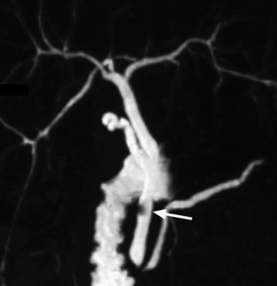
MAGNETIC RESONANCE CHOLANGIOPANCREATOGRAPHY (MRCP)
Technique
 Stationary water appears as areas of high SI and adjacent soft tissue is low SI (therefore it is not reliant on contrast excretion and can be used in jaundiced patients)
 Fasting reduces any unwanted signal from the adjacent intestine
 Breath-hold or non-breath-hold (respiratory triggered) imaging
Normal anatomy
 extrahepatic ducts ≤ 7mm (CBD up to 10mm post cholecystectomy)
  pancreatic duct ≤ 3mm
  accessory pancreatic duct in 45%
 separate drainage of segment I
 anterior (30%)
  posterior (20%)
 drainage of the right anterior or posterior duct into the left hepatic duct
  a triple confluence at the hilum
Imaging pitfalls
 MIP reformats can also over- and underestimate strictures
 a contracted sphincter mimicking an impacted stone
 flow phenomena (central signal void)
  debris
  haemorrhage
 susceptibility artefact from surgical clips
HEPATOBILIARY SCINTIGRAPHY
 It is injected intravenously with serial images obtained over 2–4 h (it requires near-normal bilirubin levels)
ENDOSCOPIC RETROGRADE CHOLANGIOPANCREATOGRAPHY (ERCP)
 It also allows for: biopsy
  brushings
  sphincterotomy
  stone extraction
  biliary stenting
  biliary stricture dilatation
CHOLELITHIASIS AND CHOLEDOCHOLITHIASIS
CHOLELITHIASIS
DEFINITION
 there is a small lifetime risk of developing a gallbladder carcinoma
 pigment stones composed of calcium bilirubinate (up to 30%)
PEARLS
 a non-fasting state
  an abnormal gallbladder position
  emphysematous cholecystitis
  a gallbladder full of stones
 it is commonly see with fasting states, critically ill patients, pregnancy, and in those patients receiving total parenteral nutrition
  it resolves spontaneously in 50% of cases
CHOLECYSTITIS
ACUTE CALCULOUS CHOLECYSTITIS
Radiology Key
Fastest Radiology Insight Engine

 the assessment of bile leaks and biliary communication with cysts
  the demonstration of segmental obstruction

 a contracted gallbladder (e.g. following a recent meal)
 it can also allow fine-needle aspiration cytology to be performed
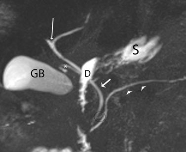
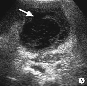
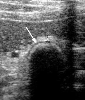
 larger stones tend to be laminated
 gallstones appear as echogenic foci which cast acoustic shadows
  stone mobility is frequently demonstrated (unless it is impacted at the neck)
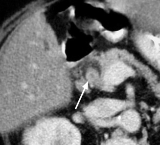
 these are hypodense, hyperdense or of mixed density
 small gallstones can be difficult to detect if they lie within any sludge


 primary sclerosing cholangitis
  recurrent pyogenic cholangitis
  Caroli’s disease
 obstructive jaundice
  pancreatitis
 a duct diameter < 4mm carries a high negative predictive value for choledocholithiasis (regardless of the gallbladder status)


 specificity > 95%)
 it can diagnose stones that are <5mm in diameter
  its main weakness is its reliance on a near-normal serum bilirubin
 this has a high sensitivity (up to 94%) and specificity (99%)
  its quality is independent of the serum bilirubin levels
 haemobilia
  flow voids
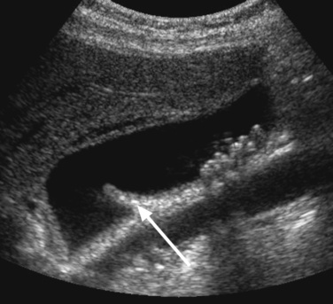
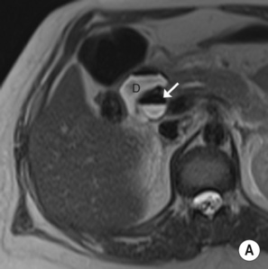
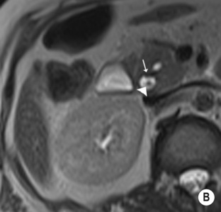
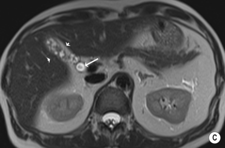
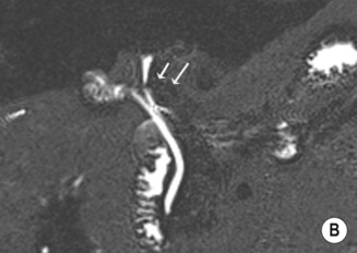
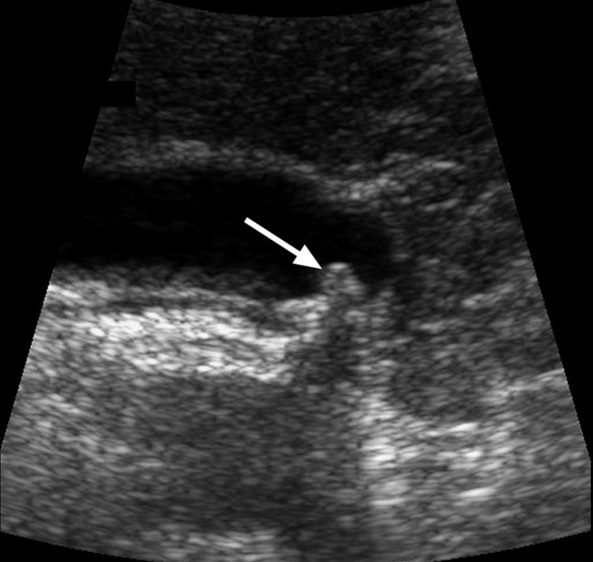
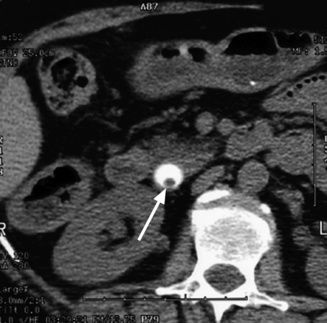
 hypoechoic line between two echogenic lines (arrow).*
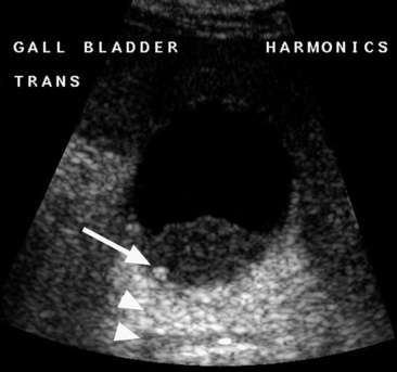
 the signs include:
 gallbladder distension (>5cm)
  pericholecystic fluid and gallbladder wall striations (± wall hyperaemia on Doppler)
  gallstones (common bile duct stones are suggested by abnormal liver function tests)
 subserosal oedema and gallbladder distension
  high-density bile
  pericholecystic fluid and inflammatory stranding within the pericholecystic fat
  variable enhancement of the gallbladder wall
 emphysematous cholecystitis
  empyema formation
 hepatitis
  pancreatitis
  gallbladder wall varices
  adenomyomatosis
  gallbladder carcinoma
 internal membranous echoes resulting from sloughed mucosa
  pericholecystic fluid
 discontinuous (±) irregular mucosal enhancement
  internal membranes (representing sloughed mucosa)
  a pericholecystic abscess
 it accounts for 1% of cases of acute cholecystitis, and has a relatively high mortality rate
 gallstones are only seen in <50% of patients
 a curvilinear brightly echogenic band with acoustic shadowing seen within a non-dependent portion of the gallbladder (representing intraluminal gas)
 this is usually found in critically ill patients
 parenteral nutrition
  AIDS
AIDS  diabetes
  chemotherapy
 gallbladder wall thickening
  echogenic contents (± sloughed membranes or mucosa)
  pericholecystic fluid
 localized gallbladder tenderness is a good predictive sign but it is difficult to assess
 intramural epithelial crypts (Rokitansky–Aschoff sinuses)
 an ejection fraction < 35% indicates gallbladder dysfunction
 it may simulate a malignancy radiologically and pathologically
 the majority have gallstones (± perforation, abscess, or fistula formation)